Cervical Instability and Vagal Compression: The Hidden Structural Driver of Dysautonomia
For a meaningful subset of dysautonomia patients, the problem is not metabolic, immune, or psychiatric. It is mechanical. The vagus nerve runs through a narrow corridor of the neck adjacent to the C1–C2 vertebrae, and structural instability in that region can directly compromise vagal signaling. A 2025 paper in Frontiers in Neurology coined the term "cervicovagopathy" to describe this — ligamentous cervical instability and dysstructure as a potential etiology for vagus nerve dysfunction (Frontiers in Neurology, 2025). It is the missing diagnosis for patients whose autonomic problems do not respond to autonomic interventions alone.
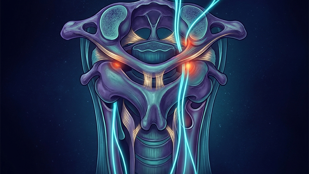
The Anatomy Matters
The vagus nerve exits the skull through the jugular foramen, descends in the carotid sheath adjacent to the internal carotid artery and internal jugular vein, and travels alongside the C1–C3 vertebrae before entering the thorax. Anything that compresses this corridor — bony malposition, ligamentous laxity, soft-tissue contracture, or vascular compromise — can affect vagal output. Upper cervical malrotations from instability are particularly impactful because the nerve is so superficial and exposed at that level.
What Cervicovagopathy Looks Like
The clinical phenotype overlaps heavily with classic dysautonomia: palpitations, dizziness on standing, GI dysmotility, fatigue, and brain fog. The distinguishing features are:
- A history of head or neck trauma — whiplash, concussion, sports injury, or surgical positioning
- Symptoms worsened by specific head positions (chin tuck, extension, rotation)
- Visible head-on-neck postural asymmetry
- Disproportionate suboccipital and upper cervical muscular tension
- Hypermobility — particularly hEDS or hypermobility spectrum disorder
- Symptoms that have failed to respond to autonomic interventions alone
Disruption of vagal signaling at this level can contribute to dizziness, palpitations, digestive issues, throat tightness, and fatigue — exactly the cluster patients describe.
Why It Gets Missed
Standard imaging — supine MRI, neutral X-ray — frequently fails to reveal upper cervical instability because the instability is dynamic and positional. The patient's neck looks normal lying down. Standing or stressed views, dynamic flexion-extension imaging, and digital motion X-ray (DMX) can reveal abnormalities invisible to conventional protocols.
Compounding the problem: most clinicians are not trained to think structurally about dysautonomia. The default assumption is that autonomic symptoms have autonomic causes — and the cervical contribution is overlooked.
The hEDS Connection
Hypermobile EDS is overrepresented in this population for an obvious reason: defective collagen makes the cervical ligaments less reliable. The same connective tissue that allows joint hypermobility allows the upper cervical spine to drift in ways that compromise the vagus. This is one of the mechanisms behind the hEDS-POTS overlap, and it explains why some EDS patients improve dramatically with structural interventions that would do nothing for an otherwise similar non-hypermobile patient.
A Diagnostic Workup for Suspected Cervicovagopathy
- Targeted history. Trauma, hypermobility, positional symptoms.
- Postural assessment. Forward head, head tilt, cervical asymmetry.
- Provocative testing. Symptom changes with cervical positioning.
- Upright cervical imaging. Standing flexion-extension X-rays, DMX, or upright MRI where available.
- Specialist referral. A clinician trained in upper cervical biomechanics — chiropractic, osteopathic, or neurosurgical depending on severity.
Treatment Layers
- Conservative care first. Prolotherapy, cervical-specific physical therapy, postural retraining, and avoidance of provocative positions.
- Co-treat the autonomic system. Vagal restoration practices, HRV training, hydration and salt for orthostatic stability. Structural and autonomic care are complementary, not competitive.
- Surgical intervention is rare and last-resort. Reserved for severe, demonstrable instability that has failed conservative care.
Clinical takeaway: For a meaningful subset of dysautonomia patients, the autonomic problem has a structural cause. Cervicovagopathy is a real clinical entity. If a patient with the right history is not improving on autonomic interventions, the neck deserves a careful look.
References & Further Reading
- Cervicovagopathy: ligamentous cervical instability as a cause of vagus nerve dysfunction. Frontiers in Neurology, 2025. Read
- Cervicovagopathy — PMC version. PMC 12263383. Read
- Ligamentous cervical instability — forward head, fluid flow. Frontiers in Neurology, 2024. Read
- A guide to identify cervical autonomic dysfunctions. PMC 10201454. Read
Have a question?
Have a question about something specific? Send us a message.
Visit VagusSkool.com/contact — we'll try to get back to you within 24 hours.