The Vagus Nerve, Sleep Architecture, Stress Recovery, and Resting Heart Rate
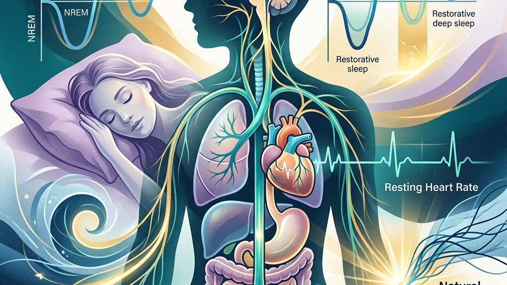
The vagus nerve serves as a primary conduit between the brainstem and major organs, shaping how the body transitions between states of alertness and restoration. Its activity influences the depth of nighttime rest, the speed of return to calm after pressure, and the baseline tempo of the heartbeat at rest. Understanding these connections offers a clearer picture of why some people experience fragmented sleep, prolonged tension, or elevated resting heart rates even when no acute threat is present.
This article examines the anatomical reach of the vagus nerve, its role in parasympathetic regulation, and the specific ways its tone intersects with sleep continuity, recovery from stress, and cardiac rhythm. Readers will encounter the underlying physiology, patterns commonly reported in daily life, and the current state of evidence drawn from established sources. The discussion remains grounded in mechanisms rather than guarantees of change.
How the vagus nerve works
The vagus nerve, designated as cranial nerve ten, originates in the medulla oblongata and extends long branches through the neck, chest, and abdomen. It carries both sensory information from visceral organs back to the brain and motor signals that slow heart rate, stimulate digestion, and modulate inflammation. Roughly eighty percent of its fibers are afferent, meaning they primarily relay data upward rather than issuing commands downward. These ascending fibers travel from stretch receptors in the lungs, pressure sensors in the aortic arch, and chemoreceptors along the gastrointestinal tract, delivering continuous updates about organ status that the brainstem integrates into moment-to-moment adjustments of autonomic outflow.
Its parasympathetic functions form the core of the “rest and digest” system. When vagal outflow increases, acetylcholine release at cardiac synapses lengthens the interval between heartbeats, while parallel signals to the lungs and gut promote slower respiration and enhanced motility. This same nerve participates in the gut-brain axis by transmitting microbial and mechanical signals from the intestines that can influence mood and arousal thresholds. In everyday terms, the post-meal sensation of drowsiness after a balanced lunch reflects acetylcholine-mediated increases in intestinal blood flow and reduced sympathetic drive, allowing digestive processes to proceed without competing cardiovascular demands. Conversely, when afferent signals indicate gut distension or microbial imbalance, the brain may raise arousal thresholds, producing the restless alertness sometimes felt after an unusually heavy or unfamiliar evening meal.
Heart-rate variability serves as a practical window into vagal tone. Greater beat-to-beat fluctuation, especially in the high-frequency band linked to respiration, reflects stronger parasympathetic modulation. Lower variability often corresponds to reduced vagal influence and a more rigid autonomic state. These measurements remain indirect yet widely used markers in both research and clinical observation. For instance, a person who notices their pulse quicken sharply with a simple change from sitting to standing may be observing a narrower range of vagal buffering, whereas someone whose heart rate drifts smoothly with each breath demonstrates more flexible parasympathetic engagement during ordinary postural shifts.
Sleep and Vagal Tone
During the descent into non-REM sleep, vagal activity rises and helps stabilize slower brain-wave patterns. The nerve’s cardiac and respiratory branches contribute to the progressive slowing of heart rate and the lengthening of exhalation that characterize deeper sleep stages. When vagal tone is adequate, transitions between light and slow-wave sleep tend to occur with fewer micro-arousals, supporting the consolidation of restorative processes that occur overnight. This stabilization occurs partly because vagal afferents dampen incoming sensory traffic from the viscera, reducing the likelihood that minor intestinal contractions or slight blood-pressure fluctuations will register as arousing events at the cortical level.
People frequently notice that nights of higher vagal engagement feel subjectively deeper and leave them less reactive the following day. Conversely, evenings marked by sustained sympathetic dominance can produce lighter sleep, more frequent awakenings, and a sense that rest was incomplete despite adequate time in bed. These patterns align with the nerve’s role in dampening sensory throughput from the body to the cortex during sleep. A concrete illustration is the difference between falling asleep after an evening walk versus after an intense work call: the former often permits longer exhalations that recruit vagal cardiac fibers, while the latter leaves residual sympathetic tone that shortens exhalations and increases the probability of shifting back into lighter stages.
Respiratory sinus arrhythmia, the natural acceleration of heart rate on inhalation and deceleration on exhalation, becomes more pronounced under strong vagal influence and is often preserved during healthy sleep cycles. Disruption of this rhythm through shallow or irregular breathing can fragment sleep architecture even when external conditions appear favorable. Over successive nights, such fragmentation may accumulate into daytime fatigue without an obvious external cause. Individuals who habitually breathe from the upper chest during the day may carry that pattern into the night, reducing the amplitude of respiratory sinus arrhythmia and thereby limiting the vagal support that normally accompanies slow-wave sleep.
Research on sleep-disordered breathing further illustrates vagal involvement. Altered vagal signaling can coincide with changes in upper-airway muscle tone and ventilatory control, creating a bidirectional relationship between breathing stability and autonomic balance throughout the night. Individuals sometimes observe that improving daytime breathing patterns coincides with fewer nighttime disruptions, though the direction of influence varies. For example, someone who practices slower diaphragmatic breathing in the late afternoon may notice reduced snoring or fewer positional awakenings, reflecting improved coordination between vagal motor fibers to the larynx and the central respiratory rhythm generators.
Stress Recovery and Vagal Pathways
After an acute stressor ends, the vagus nerve facilitates the return to baseline by inhibiting sympathetic outflow and lowering inflammatory signaling. This “vagal brake” operates through direct projections to the heart and through anti-inflammatory pathways that involve the spleen and other immune tissues. Efficient engagement of these routes shortens the duration of elevated heart rate, muscle tension, and circulating stress mediators. The brake engages when baroreceptors detect stable blood pressure and send afferent signals that prompt the nucleus tractus solitarius to increase vagal efferent traffic, rapidly lengthening cardiac intervals and reducing peripheral vascular tone.
Many people describe a lingering sense of activation long after the original pressure has passed. When vagal tone is lower, the system may remain biased toward sympathetic readiness, producing sustained shallow breathing, digestive slowdown, or a feeling of being “wired but tired.” Repeated cycles of incomplete recovery can gradually narrow the window of tolerance for subsequent stressors. A typical example is the professional who finishes a deadline at 9 p.m. and then finds the digestive system unresponsive to a late dinner; reduced vagal motility leaves food sitting longer in the stomach, which in turn sends afferent signals that maintain a low level of cortical arousal.
The nerve’s sensory fibers also convey information about visceral state back to regulatory centers in the brainstem and forebrain. This feedback loop allows the brain to update its assessment of safety or threat based on bodily signals. When vagal traffic carries calmer visceral information, the brain is more likely to down-regulate defensive responses, supporting quicker psychological as well as physiological settling. After a difficult conversation, for instance, the difference between a person who notices gradual softening of abdominal tension and one who remains unaware of visceral signals often tracks with how readily the brain accepts the new, lower-threat appraisal.
Chronic low-grade stress can reduce the dynamic range of vagal responses, making it harder for the system to shift gears even when environmental demands decrease. Observations in daily life include difficulty falling asleep after evening work demands or an extended period of gastrointestinal unease following social strain. These experiences reflect the same underlying limitation in parasympathetic rebound capacity. Over weeks, the pattern may appear as a consistently smaller difference between daytime and nighttime heart-rate variability readings on a wearable device, indicating that the vagal brake is available but engages more slowly.
Resting Heart Rate and the Vagal Brake
At rest, the vagus nerve supplies continuous inhibitory tone to the sinoatrial node, keeping heart rate below its intrinsic pacemaker rate. This tonic influence produces the characteristic beat-to-beat variability that buffers against sudden accelerations. When vagal outflow is robust, resting heart rate tends to sit in a lower range and shows greater responsiveness to respiratory phase. The brake is not a fixed clamp; it modulates dynamically with each breath, releasing slightly on inhalation and strengthening on exhalation, which explains why resting heart rate can vary by several beats per minute within a single minute of quiet sitting.
Individuals often notice that their morning pulse feels steadier or slightly slower on days when prior sleep and daytime calm have been adequate. Conversely, periods of sustained pressure or poor recovery can manifest as a persistently elevated resting rate that does not drop as readily with simple relaxation. These shifts occur without changes in fitness level and reflect autonomic balance rather than structural cardiac alteration. Someone who measures their pulse while still in bed may observe that the same body position yields different readings depending on whether the preceding evening included a period of quiet reading or continued screen-based work.
The vagus nerve’s effect on the heart is not uniform; it interacts with sympathetic inputs and with baroreceptor reflexes that sense blood-pressure fluctuations. Stronger vagal modulation enhances the efficiency of these reflexes, allowing heart rate to adjust smoothly to postural changes or mild exertion. Reduced modulation can produce a flatter response curve, so that heart rate remains elevated longer after minor activity. For example, climbing a single flight of stairs may return a person with higher vagal tone to baseline within thirty seconds, whereas the same exertion may require two minutes for heart rate to settle when vagal influence is lower.
Over time, the cumulative impact of lower vagal tone on resting cardiac parameters may appear in metrics such as reduced heart-rate variability during quiet sitting or standing. People sometimes track these patterns through wearable devices and observe that days with higher variability coincide with lower average resting rates and a subjective sense of greater ease in the chest and breath. This correspondence arises because the same vagal fibers that slow the sinoatrial node also refine the timing of each beat relative to the respiratory cycle, producing both the numerical variability and the felt sense of steadiness.
What the research shows
Studies examining heart-rate variability consistently link higher cardiac vagal tone to improved autonomic flexibility across multiple systems. research on heart-rate variability and cardiac vagal tone describes how respiratory-linked fluctuations serve as a reliable index of parasympathetic regulation and its relationship to both cardiovascular and emotional adaptability. These findings provide a measurable bridge between nerve activity and observable resting-state physiology.
Investigations into sleep have identified associations between vagal stimulation parameters and changes in sleep continuity as well as breathing stability. research on vagus nerve stimulation, sleep-disordered breathing, and sleep quality outlines how modulation of vagal pathways can intersect with respiratory control during sleep, offering mechanistic context for why some individuals experience fewer arousals when autonomic balance improves.
Additional work on the gut-brain axis highlights the vagus nerve’s role in transmitting signals that influence central regulatory centers involved in arousal and recovery. research on the vagus nerve as modulator of the brain–gut axis details bidirectional communication routes that can affect both digestive function and higher-order processes tied to stress responsiveness. Complementary cardiovascular studies further show how vagal stimulation influences heart-rate dynamics and inflammatory tone in parallel. research on vagus nerve stimulation and the cardiovascular system maps these direct cardiac effects.
Anatomical reviews confirm the extensive distribution of vagal fibers and their integration with brainstem nuclei that coordinate parasympathetic output. neuroanatomy of the vagus nerve supplies the structural foundation for understanding why interventions targeting the nerve can produce widespread effects on heart rate, respiration, and visceral tone. Clinical summaries from major medical centers similarly describe the nerve’s dual sensory-motor functions and its relevance to autonomic balance. overview of vagus nerve function and conditions
Practical ways to support your vagus nerve
Because vagal tone reflects dynamic interactions among respiratory, cardiovascular, and visceral systems, practices that gently recruit its afferent or efferent pathways can be integrated into ordinary routines without requiring specialized equipment. The following approaches emphasize low-intensity stimuli that align with the nerve’s natural response characteristics.
- Slow extended exhales performed for several minutes while seated can lengthen the phase of the respiratory cycle that most strongly recruits vagal cardiac fibers, gradually increasing heart-rate variability.
- Humming or gentle gargling engages the laryngeal and pharyngeal branches of the vagus, providing a simple mechanical stimulus that many people incorporate at the beginning or end of the day.
- Brief, tolerable cold exposure such as cool water on the face or wrists activates vagal afferents through temperature-sensitive receptors and can produce a noticeable shift in breathing depth afterward.
- Paced breathing at roughly six breaths per minute aligns with the frequency at which baroreflex and vagal systems interact most efficiently, supporting steadier resting cardiac rhythms over repeated sessions.
- Light movement that includes gentle neck and shoulder mobility maintains afferent traffic from cervical vagal branches without requiring high exertion levels.
- Consistent morning light exposure combined with a stable sleep schedule helps anchor circadian signals that in turn influence overnight vagal dominance during sleep cycles.
When to talk to a professional
Sudden or unexplained changes in resting heart rate, persistent sleep fragmentation that does not improve with basic routine adjustments, or prolonged difficulty returning to calm after ordinary stressors warrant evaluation by a qualified clinician. These signs may reflect interactions among multiple systems rather than isolated vagal function.
Individuals experiencing chest discomfort, dizziness on standing, severe gastrointestinal changes, or breathing difficulties at night should seek prompt medical assessment. Professional guidance ensures that any observed patterns receive appropriate investigation within the full clinical context.
Common questions
How quickly can vagal tone change?
Short-term shifts in heart-rate variability can appear within minutes of breathing or postural adjustments, yet longer-term stabilization of resting parameters generally unfolds over weeks of consistent practice and recovery habits.
Does age affect vagal influence?
Vagal tone tends to decline gradually with advancing age, which can contribute to reduced heart-rate variability and altered sleep depth, although individual trajectories vary widely depending on overall health and lifestyle patterns.
Can devices accurately track vagal activity?
Wearable metrics such as heart-rate variability during sleep or quiet rest provide useful approximations of vagal modulation, yet they remain indirect and are best interpreted alongside subjective experience and professional oversight when concerns arise.
Is there a single best practice for everyone?
No universal sequence exists; responses differ according to baseline autonomic state, so beginning with the least demanding options and observing personal patterns allows for more tailored adjustments over time.
The vagus nerve offers one coherent thread connecting nighttime restoration, daytime recovery, and the quiet tempo of the resting heart. Attention to its signals through steady habits can illuminate how these systems interact without replacing individualized medical care when symptoms persist or intensify.
Have a question?
Have a question about something specific? Send us a message.
Visit VagusSkool.com/contact — we'll try to get back to you within 24 hours.