The Vagus Nerve's Role in Digestive Processes and Resting Cardiac Rhythm
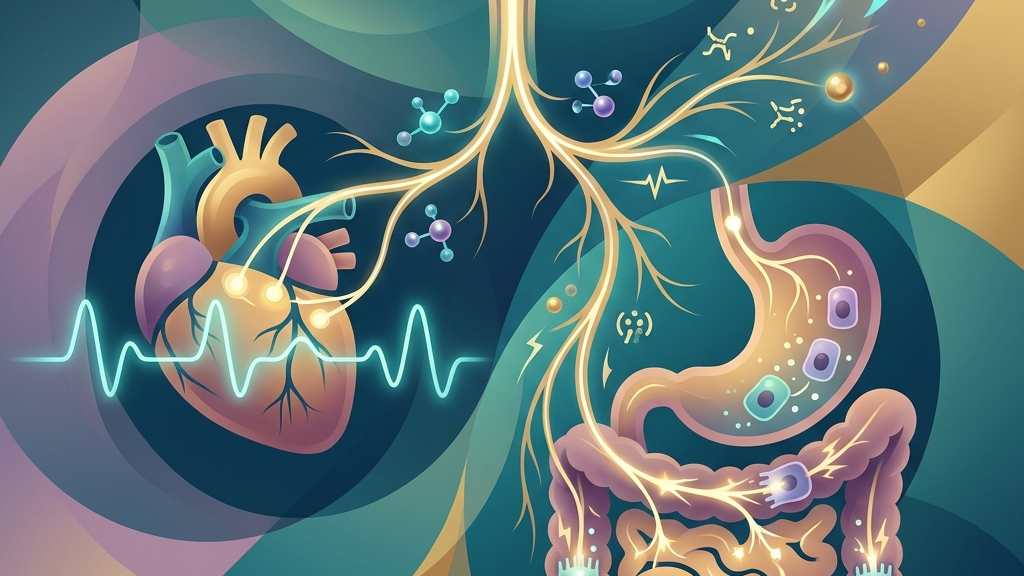
The vagus nerve serves as a primary conduit for parasympathetic signaling throughout the body, linking the brainstem to major organs involved in nutrient processing and cardiovascular stability. Its extensive reach allows bidirectional communication that can shape how efficiently food moves through the gastrointestinal tract and how steadily the heart maintains its baseline pace during periods of calm. Understanding these connections offers insight into the broader autonomic framework that supports daily physiological balance. Everyday experiences such as the gradual settling of digestion after a balanced meal or the slowing of pulse during quiet reading illustrate how this nerve integrates visceral activity with central regulation, providing a continuous stream of data that helps maintain equilibrium without conscious effort.
Readers will encounter detailed explanations of the nerve's anatomical layout, its specific contributions to digestion and heart-rate regulation, and the physiological mechanisms that underlie common observations. The discussion draws on established neuroanatomy and emerging research to clarify how vagal pathways operate without implying any direct interventions or outcomes. This foundation prepares the ground for considering supportive habits and appropriate moments to seek clinical guidance. By examining these processes at the level of cellular signaling and organ-level coordination, individuals gain a clearer picture of why certain patterns recur in daily life, such as changes in comfort after eating or shifts in perceived energy during rest.
How the Vagus Nerve Works
The vagus nerve emerges from the medulla oblongata as the tenth cranial nerve and extends the longest course of any cranial nerve, descending through the neck, thorax, and abdomen. It carries both efferent fibers that convey commands from the central nervous system to peripheral organs and afferent fibers that relay sensory information back to the brainstem. This dual traffic supports its dominant role within the parasympathetic division of the autonomic nervous system, promoting restorative functions such as slowed cardiac activity and enhanced gastrointestinal motility when the body is not under acute demand. The nerve's myelinated and unmyelinated axons allow for rapid and sustained signaling, respectively, enabling both immediate adjustments like a brief pause in swallowing and longer-term modulation of organ tone across hours.
Within the gut-brain axis, vagal afferents sample mechanical stretch, chemical composition, and microbial metabolites in the intestinal lumen, transmitting these signals to brainstem nuclei that integrate them with emotional and cognitive processing centers. Efferent projections then modulate smooth-muscle tone, glandular secretion, and local blood flow. The resulting loop allows the brain to adjust digestive priorities in response to overall physiological state while simultaneously informing higher centers about visceral conditions that may influence mood or alertness. For instance, after consuming a fiber-rich lunch, stretch receptors along the stomach wall activate specific afferent branches that reach the nucleus tractus solitarius within milliseconds, prompting refined adjustments in enzyme release that continue into the afternoon without disrupting ongoing mental tasks.
Heart-rate variability arises largely from fluctuating vagal outflow to the sinoatrial node. Respiratory sinus arrhythmia, for instance, reflects cyclic increases and decreases in vagal tone synchronized with breathing, producing measurable beat-to-beat differences. Higher variability at rest generally corresponds to greater parasympathetic dominance, whereas reduced variability can indicate a shift toward sympathetic prevalence or diminished vagal capacity. These patterns provide a noninvasive window into the dynamic balance maintained by the vagus nerve across organ systems. In practice, someone sitting quietly at a desk may notice subtle pulse changes that align with each breath cycle, reflecting how the nerve fine-tunes cardiac output to match oxygen needs during low-demand periods.
How the Vagus Nerve Supports Digestive Processes
Vagal efferents densely innervate the esophagus, stomach, pancreas, and proximal small intestine, releasing acetylcholine that promotes peristaltic waves and relaxation of sphincters at appropriate intervals. In the stomach, this innervation coordinates receptive relaxation during food intake and subsequent mixing contractions, while in the intestines it facilitates segmental propulsion and coordinated secretion of digestive enzymes and bicarbonate. Disruption of these signals can slow transit or alter secretory patterns, illustrating the nerve's direct contribution to mechanical and chemical aspects of digestion. A concrete example occurs during a relaxed evening meal: vagal activation encourages the lower esophageal sphincter to open briefly for swallowed food while maintaining tone afterward to prevent reflux, all calibrated to the volume and consistency of what was eaten.
Afferent fibers traveling within the vagus continuously monitor luminal distension, nutrient concentration, and local pH, feeding data into the nucleus tractus solitarius. From there, reflex arcs adjust gastric tone and intestinal motility within seconds, creating a feedback system that matches digestive activity to the composition and volume of incoming material. This sensory traffic also reaches hypothalamic and limbic structures, allowing visceral states to influence broader regulatory networks that govern appetite and satiety over longer timescales. When a person eats a meal containing both protein and fat, chemosensitive vagal endings detect rising amino-acid and fatty-acid levels, triggering graded reductions in gastric emptying that spread nutrient absorption evenly across the next several hours.
Many people notice that periods of heightened stress coincide with sensations of bloating, slowed gastric emptying, or irregular bowel patterns. These experiences align with a temporary withdrawal of vagal tone that favors sympathetic activation, reducing motility and altering secretion. Conversely, when parasympathetic dominance returns, individuals often report improved comfort after meals and more predictable transit, reflecting restored coordination between central commands and peripheral gut behavior. An office worker who steps away from a tense meeting for a short walk may later observe steadier digestion during dinner, as renewed vagal signaling resumes normal peristaltic sequencing.
The same afferent pathways participate in signaling satiety hormones such as cholecystokinin and glucagon-like peptide-1, which amplify vagal messages to brainstem centers that regulate meal termination. Over repeated cycles, this integration helps calibrate portion size and meal frequency to metabolic need. Subtle day-to-day variations in these signals can therefore contribute to the familiar sense that digestion feels efficient on some days and sluggish on others, even when dietary intake remains comparable. For example, after several nights of shorter sleep, the same breakfast portion may produce prolonged fullness because circadian shifts have modestly reduced vagal sensitivity to those hormonal cues.
Vagal Modulation of Resting Heart Rate
At rest, tonic vagal activity exerts a continuous braking effect on the sinoatrial node, keeping heart rate below the intrinsic pacemaker rate of approximately 100 beats per minute. This "vagal brake" operates through rapid acetylcholine release that hyperpolarizes pacemaker cells, lengthening the interval between depolarizations. Because vagal influence can be withdrawn or augmented within a single cardiac cycle, it provides fine-grained control that sympathetic pathways alone cannot achieve with comparable speed. During a quiet evening of reading, this mechanism keeps the pulse in the low seventies for most adults, adjusting smoothly when posture changes from sitting to reclining.
Respiratory modulation of this brake generates the characteristic rise in heart rate during inspiration and fall during expiration. The pattern emerges from central respiratory drive inhibiting vagal preganglionic neurons during inhalation, allowing transient sympathetic predominance, followed by rebound vagal activation on exhalation. The resulting variability in inter-beat intervals serves as a functional index of vagal capacity and overall autonomic flexibility. Someone practicing slow breathing while waiting for an appointment may observe on a wearable device that the spread between consecutive beats widens noticeably during the longer exhale phases.
Individuals frequently observe that their resting pulse feels slower and more steady after extended periods of quiet breathing or relaxation, consistent with augmented vagal outflow. Conversely, mental preoccupation or physical tension can produce a quicker, less variable pulse even without overt movement. These subjective impressions correspond to shifts in the balance between vagal and sympathetic influences on the node, illustrating how everyday psychological states register in cardiac rhythm. A student reviewing notes before an exam often experiences this quickening, which reverses once the immediate cognitive demand subsides.
Baroreflex arcs further engage the vagus to buffer blood-pressure fluctuations. When arterial pressure rises, baroreceptor afferents trigger increased vagal efferent activity that slows the heart, thereby lowering cardiac output and helping restore pressure. This reflex operates continuously at rest, contributing to the stability many people associate with feeling "settled" rather than jittery or racing. Subtle impairments in baroreflex sensitivity can therefore manifest as greater moment-to-moment fluctuations in perceived heart rhythm. Standing up quickly after prolonged sitting, for instance, briefly engages this reflex to counteract the minor pressure drop and maintain steady cerebral perfusion.
What the Research Shows
Neuroanatomical tracing studies confirm that vagal sensory neurons densely innervate the gastrointestinal tract and convey information critical to brain-gut communication, as detailed in research on vagal sensory neurons and gut-brain signaling. Complementary work examining the vagus as a modulator of the brain-gut axis highlights its role in integrating nutrient signals with central regulatory circuits, documented in studies on the vagus nerve as modulator of the brain–gut axis. These investigations map precise terminal fields within the intestinal mucosa and submucosa, revealing how individual fiber types respond selectively to mechanical versus chemical stimuli.
Cardiac investigations demonstrate that resting heart-rate variability largely reflects vagal tone, with higher variability linked to greater parasympathetic influence, according to research on heart rate variability and cardiac vagal tone. Additional findings connect vagal pathways to respiratory modulation during sleep, where altered vagal activity appears in work on vagus nerve stimulation, sleep-disordered breathing, and sleep quality. Longitudinal recordings show that the magnitude of respiratory sinus arrhythmia correlates with nightly transitions between sleep stages, underscoring the nerve's continuous role in autonomic stability.
Standard anatomical references from Cleveland Clinic descriptions of vagus nerve function and distribution and NIH StatPearls coverage of cranial nerve 10 neuroanatomy provide the foundational mapping of efferent and afferent projections that underpins these functional observations. Together the sources illustrate consistent patterns across cardiac, gastrointestinal, and central nervous system domains without establishing causal claims for any specific intervention.
Practical Ways to Support Your Vagus Nerve
- Slow, extended exhales performed for several minutes can increase the duration of vagal dominance during each respiratory cycle, as longer exhalations permit greater acetylcholine release at the sinoatrial node.
- Humming or gentle gargling engages laryngeal and pharyngeal branches of the vagus, creating mechanical stimulation that may transiently elevate vagal afferent traffic to the brainstem.
- Brief, tolerable cold exposure such as cool water on the face activates trigeminal-vagal reflexes that can produce measurable shifts in heart-rate variability within seconds.
- Paced breathing at approximately six breaths per minute aligns with the resonant frequency of the baroreflex, amplifying vagal oscillations and supporting steadier resting cardiac rhythm.
- Light rhythmic movement such as walking or rocking stimulates proprioceptive and vestibular afferents that communicate with vagal nuclei, potentially reinforcing parasympathetic tone after periods of stillness.
- Consistent morning light exposure combined with stable sleep timing helps entrain circadian regulation of autonomic balance, indirectly sustaining the conditions under which vagal outflow predominates during rest.
When to Talk to a Professional
Sudden or persistent changes in resting heart rate, such as unexplained tachycardia, bradycardia, or irregular rhythms, warrant prompt medical evaluation to rule out structural or electrical cardiac issues. Similarly, severe or rapidly worsening digestive symptoms including intractable nausea, significant abdominal pain, or abrupt alterations in bowel habits deserve clinical assessment rather than self-interpretation.
Individuals experiencing chest discomfort, dizziness, fainting, or shortness of breath alongside any perceived change in vagal function should seek immediate care, as these signs may indicate conditions requiring urgent attention. Professional guidance ensures that any observed patterns receive appropriate diagnostic context and that underlying medical factors are addressed by qualified practitioners.
Common Questions
What does the term vagal tone describe?
Vagal tone refers to the baseline level of parasympathetic activity transmitted through the vagus nerve, often inferred from heart-rate variability at rest. It indexes the degree to which vagal efferents restrain cardiac pacemaker activity and support restorative organ functions. Measurements typically involve time-domain or frequency-domain analysis of electrocardiographic recordings collected during quiet wakefulness.
Can breathing exercises influence digestion through the vagus nerve?
Slow breathing may enhance vagal outflow, which in turn can promote coordinated gastrointestinal motility and secretion via parasympathetic pathways. The effect remains indirect and varies with individual physiology and context. For example, a short session of paced breathing after lunch may coincide with more comfortable post-meal sensations in some people, though outcomes differ across meals and days.
How is heart-rate variability connected to the vagus nerve?
Beat-to-beat fluctuations in heart rate largely reflect cyclic changes in vagal inhibition of the sinoatrial node, especially those synchronized with respiration. Greater variability generally corresponds to stronger and more flexible vagal modulation. Wearable monitors capture these fluctuations as standard deviation of normal-to-normal intervals or root mean square of successive differences, providing quantitative markers of the underlying neural traffic.
Is there a single practice that reliably strengthens vagal function?
No isolated activity has been shown to produce uniform, lasting changes across populations. Multiple low-intensity behaviors may collectively support conditions favorable to vagal activity, yet individual responses differ. Factors such as age, fitness level, and concurrent health conditions contribute to this variability in observed patterns.
Does age affect vagal regulation of the heart and gut?
Research indicates gradual reductions in heart-rate variability with advancing age, consistent with diminished vagal capacity, while gastrointestinal vagal reflexes may also show altered sensitivity. These shifts remain within normal variation and require individualized clinical interpretation. Longitudinal studies document these trends across decades while emphasizing that wide inter-person differences persist at every life stage.
The interplay between vagal pathways, digestive coordination, and resting cardiac rhythm underscores the integrated nature of autonomic regulation across organ systems. Continued attention to these mechanisms, grounded in anatomical and physiological evidence, supports a measured appreciation of how the body maintains baseline stability without overinterpreting everyday fluctuations.
Have a question?
Have a question about something specific? Send us a message.
Visit VagusSkool.com/contact — we'll try to get back to you within 24 hours.