The Vagus Nerve’s Influence on Sleep Architecture, Stress Recovery, and Digestive Function
By Sterling Cooley
•
June 6, 2026
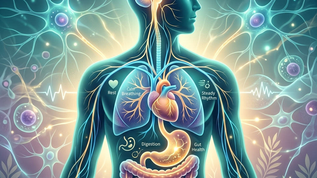
The vagus nerve serves as a primary conduit for the body’s rest-and-digest functions, linking brainstem centers with the heart, lungs, and gastrointestinal tract. Its activity shapes how readily a person transitions into deep sleep, how efficiently the cardiovascular system returns to baseline after challenge, and how smoothly nutrients are processed along the alimentary canal. Understanding these connections offers a coherent physiological framework rather than isolated tips for better rest, calmer moods, or steadier digestion. At a mechanistic level, the nerve integrates signals from multiple brainstem nuclei that continuously sample interoceptive data, allowing the body to calibrate metabolic demand against environmental safety cues. For instance, after finishing a large evening meal, vagal afferents detect gastric distension and trigger a cascade that slows cardiac output while accelerating intestinal mixing motions, creating the familiar sensation of post-dinner drowsiness. This same circuitry influences how quickly blood pressure settles following an unexpected work deadline or how consistently bowel movements occur on a predictable schedule.
This article examines the nerve’s anatomy, its role in parasympathetic regulation, and the measurable ways its tone appears in sleep continuity, post-stress heart-rate patterns, and gut motility. Evidence from neuroanatomy and clinical studies is presented without implying that any single practice guarantees improvement. Readers will encounter the mechanisms that allow the vagus nerve to dampen arousal, coordinate organ responses, and relay sensory information upward to the brain. Everyday illustrations include noticing a slower pulse while reading quietly after a stressful commute or recognizing that abdominal comfort improves on days when breathing remains unhurried during meals.
How the Vagus Nerve Works
The vagus nerve, designated cranial nerve X, originates in the medulla oblongata and descends through the neck, thorax, and abdomen, giving off branches to the larynx, heart, lungs, and most of the digestive tract. Roughly eighty percent of its fibers are afferent, carrying sensory data from visceral organs back to the brainstem; the remaining efferent fibers deliver parasympathetic commands that slow heart rate, promote bronchial constriction, and stimulate gastrointestinal secretions and motility. This bidirectional traffic forms a core part of the parasympathetic nervous system and the anatomical substrate of the gut-brain axis. The dorsal motor nucleus supplies most preganglionic efferents to thoracic and abdominal viscera, while the nucleus ambiguus contributes specialized cardio-inhibitory fibers that terminate directly on the sino-atrial node. Afferent cell bodies reside in the nodose and jugular ganglia, sending axons that terminate in the nucleus tractus solitarius, where visceral information is integrated with inputs from baroreceptors and chemoreceptors. Heart-rate variability, particularly the high-frequency component tied to respiratory sinus arrhythmia, serves as a practical index of vagal outflow to the heart. When vagal tone is robust, the interval between heartbeats lengthens on exhalation and shortens on inhalation, producing a flexible rhythm that reflects the nervous system’s capacity to modulate arousal. Lower variability often coincides with sustained sympathetic dominance, whereas higher variability tracks with efficient recovery after exertion or emotional demand. In daily life this appears as the slight acceleration felt when climbing stairs followed by a smooth deceleration once seated again, or the noticeable calming of pulse during a quiet conversation after an argument. Respiratory sinus arrhythmia arises because vagal motor neurons are phasically inhibited during inspiration by lung stretch receptors and central respiratory drive, then released during expiration, allowing acetylcholine release to hyperpolarize pacemaker cells. Because the same brainstem nuclei that regulate cardiac vagal tone also influence descending pathways to the gut and ascending pathways to higher limbic structures, changes in one domain frequently appear in others. For instance, altered vagal signaling can shift both the latency to sleep onset and the balance of sympathetic versus parasympathetic input to the enteric nervous system. These overlapping circuits explain why interventions that modestly increase vagal activity sometimes correspond with improvements across sleep, cardiovascular recovery, and digestive comfort, although individual responses vary with age, health status, and baseline autonomic balance. A person who experiences delayed gastric emptying after several nights of fragmented sleep is observing one downstream consequence of this shared circuitry.Vagal Tone and the Architecture of Restorative Sleep
During the descent into non-REM sleep, parasympathetic dominance increases and vagal cardio-motor neurons become more active, producing the characteristic rise in high-frequency heart-rate variability observed in the first sleep cycles. This shift supports the slowing of metabolic rate and the reduction of sympathetic vasoconstrictor tone that together permit deeper slow-wave activity. When vagal efferent traffic is compromised, the transition may be interrupted by micro-arousals or prolonged periods of lighter stage-two sleep, reducing the cumulative duration of slow-wave and REM stages across the night. Mechanistically, vagal activation suppresses locus coeruleus firing and reduces noradrenergic tone, allowing thalamocortical circuits to enter synchronized slow oscillations. An everyday example is the difference between falling asleep easily after a calm evening walk versus lying awake with a racing pulse following an evening of intense screen use. Sensory afferents traveling in the vagus nerve also convey information about respiratory and gastrointestinal state that helps stabilize breathing patterns during sleep. Research on sleep-disordered breathing indicates that intact vagal feedback can dampen excessive sympathetic surges that otherwise fragment sleep continuity. Individuals with lower vagal tone may therefore notice more frequent awakenings or a sense that sleep feels “light,” even when total sleep time appears adequate by conventional measures. Vagal pulmonary afferents, for instance, sense subtle changes in lung volume and relay them to the nucleus tractus solitarius, which adjusts respiratory motor output to prevent over-breathing that could trigger arousals. Over successive nights, the cumulative effect of reduced vagal modulation appears in next-day markers such as elevated resting heart rate and diminished baroreflex sensitivity. These changes can, in turn, blunt the normal evening rise in melatonin and the nocturnal dip in core body temperature, further delaying sleep onset. The relationship is bidirectional: fragmented sleep itself tends to lower vagal tone the following day, creating a self-reinforcing loop that some people experience as persistent fatigue despite extended time in bed. This loop can be observed when an individual stays up late under bright lights, then finds the next evening’s wind-down period lengthened because daytime autonomic recovery remained incomplete.The Vagus Nerve in Shifting from Stress to Physiological Recovery
Acute stress activates sympathetic pathways that accelerate heart rate and redirect blood flow toward skeletal muscle. The vagus nerve supplies the opposing “vagal brake,” rapidly increasing efferent traffic to the sino-atrial node once the stressor subsides. This quick re-engagement of parasympathetic influence produces the deceleration of heart rate and the restoration of heart-rate variability that mark successful recovery. When vagal pathways are less responsive, heart rate remains elevated longer and blood pressure returns to baseline more slowly. The brake operates through acetylcholine binding to muscarinic receptors that open potassium channels, hyperpolarizing pacemaker cells within one or two cardiac cycles. The same vagal afferents that monitor cardiac and pulmonary status also project to the nucleus tractus solitarius, which in turn modulates activity in the hypothalamus and amygdala. Enhanced vagal signaling can therefore attenuate the central stress response, lowering the likelihood that a single demanding event will trigger prolonged HPA-axis activation. People commonly report that after periods of higher vagal tone they feel able to “let go” of tension more readily, although this subjective experience tracks only loosely with laboratory indices. In practice this might appear as the ability to resume focused work after a tense phone call once breathing has returned to a slower rhythm. Repeated or sustained sympathetic dominance without adequate vagal counter-regulation may contribute to the perception of poor stress recovery, including lingering muscle tension, digestive unease, and difficulty concentrating. Because vagal motor neurons are themselves influenced by higher cortical inputs, practices that encourage slow, rhythmic breathing or gentle vocalization can transiently increase vagal outflow and thereby shorten the time required for cardiovascular and emotional settling after challenge. Prefrontal projections to the nucleus ambiguus provide one anatomical route for such top-down modulation, allowing deliberate attention to breath to influence brainstem autonomic centers.Gut-Brain Communication and Digestive Motility via the Vagus Nerve
Vagal efferent fibers stimulate gastric acid secretion, pancreatic enzyme release, and peristaltic contractions throughout the small and large intestine. These commands are coordinated with local enteric reflexes, yet the vagus provides the long-range timing that aligns digestion with periods of relative safety and low arousal. When vagal tone is reduced, gastric emptying and intestinal transit may slow, leading some individuals to notice post-meal fullness or irregular bowel patterns that lack an obvious dietary explanation. The efferent signals travel via the celiac and superior mesenteric plexuses, synapsing onto postganglionic neurons embedded in the myenteric plexus that directly innervate smooth muscle layers. Afferent vagal fibers continuously sample mechanical stretch, nutrient composition, and inflammatory signals within the gut wall, forwarding this information to the brainstem and, via relay nuclei, to insular and prefrontal cortices. This sensory stream contributes to the subjective sense of digestive comfort or discomfort and can influence mood and appetite regulation through the gut-brain axis. Studies of vagal sensory neurons demonstrate that selective activation of these fibers can alter feeding behavior and visceral pain thresholds in animal models, underscoring the nerve’s role in interoceptive awareness. A concrete illustration is the difference in perceived satiety when eating slowly enough for stretch receptors to signal fullness versus consuming the same meal rapidly. Because the vagus nerve also carries anti-inflammatory signals via the cholinergic anti-inflammatory pathway, its activity can modulate local cytokine production in the intestinal mucosa. Lower vagal tone has been associated in observational work with heightened intestinal permeability and altered microbiota composition, although causation remains under investigation. Individuals may therefore experience a cluster of symptoms—bloating, altered stool consistency, and a sense of unease after eating—that coincides with other indicators of reduced parasympathetic regulation such as low heart-rate variability. The pathway involves acetylcholine acting on alpha-7 nicotinic receptors expressed by macrophages, dampening TNF-alpha release within the lamina propria.What the Research Shows
Neuroanatomical tracing studies confirm that the vagus nerve constitutes the principal parasympathetic supply to thoracic and abdominal viscera, with dense projections to the heart and gastrointestinal tract. detailed mapping of vagal motor and sensory nuclei illustrates how a single cranial nerve can coordinate cardiac slowing, respiratory patterning, and enteric motility. Complementary physiological recordings show that respiratory sinus arrhythmia amplitude, a marker of vagal cardio-motor output, reliably tracks changes in behavioral state from wakefulness to sleep. Clinical investigations of vagal pathways during sleep document that higher nocturnal heart-rate variability predicts longer durations of slow-wave sleep and fewer respiratory events. longitudinal data on vagus nerve stimulation and sleep quality further indicate that augmenting vagal signaling can reduce the frequency of apneas and improve subjective sleep depth in selected populations. These findings align with earlier observations that cardiac vagal tone, indexed by heart-rate variability metrics, declines with aging and with chronic stress exposure. In the domain of digestive function, tracing and lesion studies establish that vagal afferents are required for normal nutrient-induced suppression of appetite and for coordinated gastric motility. reviews of vagal modulation of the brain-gut axis synthesize evidence that vagal integrity influences both central appetite circuits and peripheral inflammatory tone within the gut. Additional work on vagal sensory neurons highlights their role in transmitting microbial and mechanical signals that shape visceral perception. mechanistic studies of gut-brain signaling demonstrate that selective stimulation of vagal afferents can alter feeding patterns and pain thresholds, supporting the nerve’s position as a key bidirectional conduit. Cardiovascular recovery after stress likewise depends on vagal reactivation. analyses of heart-rate variability and cardiac vagal tone show that rapid restoration of high-frequency power after laboratory stressors correlates with lower baseline sympathetic tone and better self-reported recovery. These physiological patterns are consistent with the broader anatomical description of vagal innervation provided by major clinical references. comprehensive overviews of vagal anatomy and function emphasize that the nerve’s extensive reach explains its simultaneous influence on heart rate, respiration, and gastrointestinal activity.Practical Ways to Support Your Vagus Nerve
Slow, rhythmic breathing and other low-intensity activities can engage vagal pathways through well-characterized sensory and motor routes. The following list summarizes several approaches that have been examined in physiological studies.- Slow, extended exhales performed for several minutes while seated can increase respiratory sinus arrhythmia and thereby raise momentary vagal outflow to the heart.
- Humming or gentle gargling with water engages laryngeal branches of the vagus and may produce a brief rise in heart-rate variability that some people notice as a calmer sensation in the chest and throat.
- Brief, tolerable cold exposure such as finishing a shower with cool water on the neck and face can activate vagal afferents and support a subsequent drop in heart rate once the stimulus ends.
- Paced breathing at approximately six breaths per minute, with equal or slightly longer exhales, has been shown in multiple studies to enhance high-frequency heart-rate variability within a single session.
- Light rhythmic movement such as walking at a comfortable pace allows natural coordination between respiration and stride that can modestly elevate vagal tone without requiring intense effort.
- Consistent morning light exposure within an hour of waking helps stabilize circadian rhythms that in turn support the nocturnal rise in vagal activity necessary for deeper sleep.
When to Talk to a Professional
Sudden or severe changes in sleep continuity, heart rhythm, or digestive function warrant prompt medical evaluation, because these symptoms can reflect conditions unrelated to vagal tone. Persistent chest pain, unexplained weight loss, blood in the stool, or breathing difficulties during sleep require assessment by a qualified clinician before any lifestyle approach is considered. Individuals who experience fainting, marked dizziness on standing, or rapidly worsening gastrointestinal symptoms should seek care without delay. A healthcare provider can determine whether autonomic testing, sleep studies, or gastrointestinal evaluation is appropriate and can rule out structural or inflammatory disorders that require specific medical management.Common Questions
Does stimulating the vagus nerve guarantee better sleep?
No single intervention ensures improved sleep architecture; vagal tone is one of several physiological factors that influence sleep continuity and depth, and responses vary widely among individuals.Can heart-rate variability be measured reliably at home?
Consumer devices provide approximate estimates, yet clinical interpretation of heart-rate variability requires standardized recording conditions and consideration of age, medications, and health status.Is there a quick daily practice that reliably raises vagal tone?
Slow breathing at roughly six breaths per minute can produce measurable increases in high-frequency heart-rate variability within minutes, although the magnitude and duration of the effect differ from person to person.Do digestive symptoms always indicate low vagal tone?
Digestive comfort depends on many variables including diet, microbiota, motility disorders, and inflammation; vagal signaling is one contributing pathway rather than the sole determinant.Are there risks associated with practices that aim to increase vagal activity?
Most gentle breathing or vocalization exercises are low-risk for healthy adults, yet anyone with cardiovascular or respiratory conditions should consult a clinician before beginning new routines. The vagus nerve offers a unifying physiological thread that connects the regulation of nighttime restoration, daytime recovery from challenge, and the steady processing of nutrients. Its bidirectional traffic between brainstem and viscera underscores why modest, repeated shifts in breathing rhythm or daily light exposure can correspond with changes that people notice across multiple bodily systems, even while individual outcomes remain variable and require professional guidance when symptoms are severe.Have a question?
Have a question about something specific? Send us a message.
Visit VagusSkool.com/contact — we'll try to get back to you within 24 hours.