The Vagus Nerve: How It Shapes Stress Recovery, Digestion, and Breath
The vagus nerve serves as a primary communication highway between the brain and many internal organs, influencing how the body returns to equilibrium after challenges. Understanding its contributions to stress recovery, digestive processes, and respiratory rhythms offers a clearer picture of nervous-system regulation. This article explores the underlying anatomy, physiological mechanisms, observable patterns, and current evidence without promising specific outcomes.
Readers will encounter detailed explanations of the nerve’s parasympathetic functions, its bidirectional signaling along the gut-brain axis, and measurable indicators such as heart-rate variability. Separate sections examine each focus area in depth, followed by research summaries drawn from established sources, practical considerations, and guidance on professional consultation. The goal is to present mechanisms and nuances that support informed curiosity about bodily regulation.
How the vagus nerve works
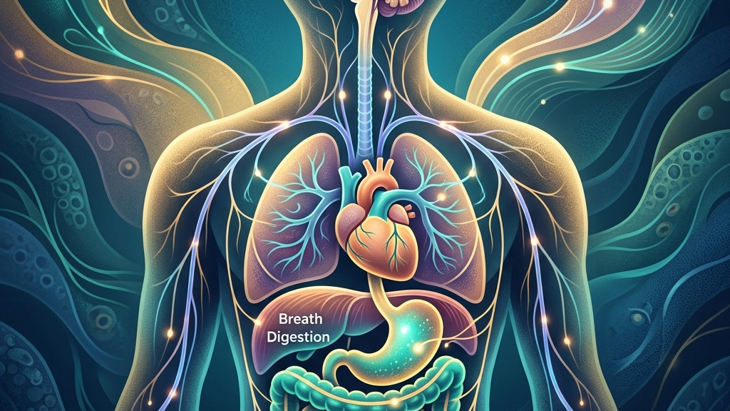
The vagus nerve, designated as cranial nerve X, emerges from the medulla oblongata and extends through the neck, chest, and abdomen, innervating structures including the heart, lungs, and gastrointestinal tract. Its parasympathetic fibers promote restorative functions by releasing acetylcholine at target organs, which slows heart rate, supports glandular secretions, and modulates smooth-muscle activity. This contrasts with sympathetic activation, creating a dynamic balance that favors recovery when vagal influence predominates. Acetylcholine binds to muscarinic receptors on cardiac pacemaker cells, hyperpolarizing the membrane and extending the time between action potentials; the same transmitter acts on enteric neurons to coordinate peristaltic waves and on pancreatic acinar cells to increase enzyme release. Because the nerve travels alongside major blood vessels and through multiple body cavities, small mechanical or inflammatory changes along its course can alter signal fidelity in localized segments without affecting the entire pathway equally.
Afferent fibers, which constitute the majority of the nerve’s axons, carry sensory information from visceral organs back to the brainstem, enabling continuous monitoring of internal states. Efferent fibers then convey regulatory signals outward. This two-way traffic underpins the gut-brain axis, allowing intestinal conditions to influence mood-related circuits and vice versa through vagal pathways. Research on vagal sensory neurons highlights their role in transmitting nutrient and inflammatory signals that shape central nervous-system responses. For example, stretch receptors in the stomach wall fire in proportion to meal volume, while chemosensitive endings detect short-chain fatty acids produced by microbial fermentation; these distinct afferent populations converge on the nucleus tractus solitarius, where they are integrated with descending cortical input before influencing hypothalamic and limbic targets. The result is a continuously updated map of visceral status that can bias attention, motivation, and autonomic set-points.
Heart-rate variability (HRV) serves as one accessible window into vagal tone. Higher variability between heartbeats often reflects stronger parasympathetic modulation, particularly via the vagus nerve’s influence on the sinoatrial node. Lower baseline variability may correspond to sustained sympathetic dominance. These patterns arise from respiratory sinus arrhythmia, in which heart rate naturally accelerates during inhalation and decelerates during exhalation under vagal control. The magnitude of this oscillation depends on both the depth of tidal volume and the prevailing level of vagal efferent traffic; when vagal tone is robust, the difference between peak inspiratory and peak expiratory intervals can exceed 100 milliseconds, whereas reduced tone compresses that range. Because HRV also incorporates slower oscillations from baroreflex and thermoregulatory loops, single-session readings require contextual interpretation rather than isolated comparison.
Recovering from Stress and the Role of Vagal Activity
Stress activates sympathetic pathways that increase heart rate, redirect blood flow, and heighten alertness. The vagus nerve contributes to counterbalancing these changes by engaging parasympathetic circuits that lower heart rate and promote visceral relaxation. When vagal outflow increases after a stressor, inflammatory cytokine production can be tempered through the cholinergic anti-inflammatory pathway, allowing physiological systems to recalibrate. This process depends on intact afferent feedback from organs that signal when conditions have stabilized. The pathway begins with vagal afferents detecting cytokine levels or tissue distension; signals reach the nucleus tractus solitarius, which activates efferent vagal neurons that release acetylcholine onto splenic macrophages, thereby suppressing TNF-α and IL-6 transcription via α7 nicotinic receptors. The timing of this anti-inflammatory brake is therefore tied to the same afferent–efferent loop that governs cardiac and gastrointestinal recovery.
Mechanistically, vagal stimulation enhances activity in the nucleus tractus solitarius, which then modulates higher centers involved in emotional appraisal. This feedback loop may reduce the duration of elevated cortisol and sympathetic tone. Individuals sometimes observe a gradual return of steady breathing, warmer extremities, and reduced muscle tension as vagal influence strengthens. These shifts are not instantaneous and vary with overall nervous-system adaptability. For instance, after an unexpected work deadline, the initial sympathetic surge may persist for 20–40 minutes even once the threat has passed; only when vagal afferents register normalized gastric motility and cardiac filling pressures does the nucleus tractus solitarius fully re-engage inhibitory projections to the amygdala and locus coeruleus.
Heart-rate variability measurements often rise during successful stress recovery, reflecting restored vagal brake function on the heart. The vagal brake refers to tonic parasympathetic inhibition that can be released quickly during threat and reapplied afterward. When this brake operates effectively, transitions between activation and rest occur more smoothly. Some people notice improved ability to shift attention away from ruminative thoughts once vagal tone rebounds. The brake’s effectiveness can be observed in everyday settings such as the difference between heart-rate recovery after climbing stairs versus after an emotionally charged conversation; the former recovers largely through baroreflex mechanisms, while the latter additionally requires vagal integration of prefrontal appraisal circuits.
Prolonged or repeated stress exposure can blunt vagal responsiveness, leading to slower return to baseline after challenges. This attenuation appears in both cardiac and gastrointestinal domains, where recovery of motility and secretion may lag. The interplay between stress history and current vagal capacity underscores why individual differences in recovery speed exist even under similar conditions. Chronic elevation of glucocorticoids down-regulates muscarinic receptor sensitivity in the sinoatrial node and reduces vagal efferent fiber excitability, producing a narrower dynamic range for respiratory sinus arrhythmia that can persist for days after the stressor subsides.
Digestive Function and Vagus Nerve Signaling
The vagus nerve supplies parasympathetic innervation to the esophagus, stomach, small intestine, and proximal colon, influencing motility, sphincter tone, and secretory activity. Acetylcholine release stimulates gastric contractions and pancreatic enzyme output while also promoting mucosal blood flow. These actions support the mechanical and chemical breakdown of food, yet they remain responsive to central states via descending vagal pathways. During a typical mixed meal, vagal efferents first increase fundic tone to accommodate volume, then shift to antral peristalsis once chyme reaches a threshold acidity; simultaneously, pancreatic vagal branches trigger bicarbonate and enzyme secretion timed to duodenal nutrient arrival. Central modulation occurs when prefrontal or limbic activity alters vagal motor nucleus output, which explains why acute worry can delay gastric emptying even when the meal composition is unchanged.
Afferent vagal fibers from the gut convey mechanical stretch, nutrient composition, and microbial metabolite information to the brainstem. This sensory stream participates in satiety signaling and can modulate reward circuits, illustrating the gut-brain axis in action. When vagal communication is efficient, digestive processes tend to proceed in coordinated waves rather than erratic patterns. Disruptions in this signaling may coincide with sensations of bloating or irregular transit that lack an obvious dietary trigger. For example, after eating a high-fiber meal, mechanoreceptors in the ileum signal distension that normally triggers the ileal brake; if vagal afferents are less responsive, the brake engages later, allowing more rapid transit and subsequent lower-bowel discomfort hours afterward.
Research on the vagus nerve as modulator of the brain–gut axis shows that vagal integrity affects both ascending sensory traffic and descending regulatory commands. For instance, vagal pathways help synchronize gastric migrating motor complexes during fasting periods. People sometimes report steadier appetite cues and fewer episodes of postprandial discomfort when overall vagal tone appears higher, though such observations remain subjective and multifactorial. Migrating motor complexes are phase-III contractions that sweep residual contents aborally; their vagal dependence is evident in the fact that they are abolished by vagotomy yet persist, albeit at lower amplitude, when only the enteric nervous system remains intact.
Anti-inflammatory effects mediated by vagal efferents also extend to the intestinal mucosa, where acetylcholine can dampen local immune activation. This mechanism may influence barrier function and motility indirectly. Because the gut contains its own enteric nervous system that interacts extensively with vagal terminals, the nerve serves more as a modulator than sole controller of digestion. Individual variability in microbiome composition further shapes the signals traveling along these fibers. Short-chain fatty acids produced by certain bacterial taxa activate vagal chemoreceptors that increase oxytocin release in the hypothalamus, illustrating one route by which microbial metabolites can bias social and feeding behaviors through the same nerve that controls motility.
Breath Patterns and Their Connection to Vagal Tone
Respiratory control and vagal activity intersect at the level of the brainstem, where vagal afferents from pulmonary stretch receptors contribute to the Hering-Breuer reflex that terminates inspiration. During exhalation, vagal cardio-inhibitory neurons become more active, producing the characteristic slowing of heart rate known as respiratory sinus arrhythmia. This coupling allows breath rhythm to serve as a lever for modulating cardiac vagal tone. The Hering-Breuer reflex prevents over-inflation by inhibiting medullary inspiratory neurons once lung volume exceeds a threshold; simultaneously, the same vagal afferents excite cardio-inhibitory neurons in the nucleus ambiguus, lengthening the subsequent R-R interval. The net effect is that each full respiratory cycle produces a measurable oscillation whose amplitude scales with both tidal volume and baseline vagal tone.
Extended or slower exhalations increase the duration of parasympathetic dominance within each respiratory cycle, often elevating short-term HRV. The mechanical act of lengthening the exhale stretches baroreceptors and lung tissue, amplifying afferent traffic that reinforces vagal outflow. Individuals may notice a subjective sense of settling or reduced intrathoracic tension when exhalations are deliberately prolonged, reflecting this physiological shift. In practice, extending the exhale from a habitual three-second duration to six seconds doubles the window during which vagal cardio-inhibitory neurons remain active, producing a larger beat-to-beat swing that can be observed on a simple pulse-oximeter waveform.
Vagal sensory neurons also monitor airway and lung status, feeding information that can alter both respiratory drive and autonomic balance. In states of heightened vigilance, breathing tends to become rapid and shallow, diminishing the oscillatory vagal input tied to full tidal volumes. Restoring slower rhythms can therefore re-engage the vagal brake on the heart and promote a broader parasympathetic tone across thoracic organs. Shallow breathing reduces phasic activation of pulmonary stretch receptors, which in turn decreases excitatory drive to the nucleus ambiguus; the resulting drop in vagal outflow allows heart rate to rise and remain elevated even when no external threat is present.
Because breathing is one of the few autonomic functions under partial voluntary control, it provides a direct route for influencing vagal activity without requiring external devices. The relationship remains bidirectional: improved vagal tone can stabilize respiratory patterns during rest, while deliberate breathing practices can transiently boost vagal metrics. These effects depend on consistency and individual baseline physiology rather than isolated sessions. Over successive days, repeated practice at resonance frequencies strengthens synaptic efficacy within the nucleus tractus solitarius–nucleus ambiguus circuit, widening the range of respiratory rates that still produce measurable HRV increases.
What the research shows
Studies examining heart-rate variability consistently link higher vagal tone to more flexible cardiovascular responses, as detailed in research on heart-rate variability and cardiac vagal tone. This body of work underscores how respiratory sinus arrhythmia serves as a proxy for parasympathetic capacity without implying diagnostic thresholds for any individual.
Investigations into the gut-brain axis demonstrate that vagal pathways transmit microbial and nutrient signals capable of altering brainstem and limbic activity, according to reviews on the vagus nerve as modulator of the brain–gut axis. Complementary findings in research on vagal sensory neurons and gut–brain signaling clarify how specific afferent populations encode mechanical and chemical information from the intestine.
Additional evidence connects vagal stimulation approaches to changes in sleep architecture and breathing stability during rest, as reported in studies of vagus nerve stimulation, sleep-disordered breathing, and sleep quality. Anatomical descriptions in NIH StatPearls on cranial nerve 10 and Cleveland Clinic materials on vagus nerve function provide foundational maps of fiber distribution that support these functional observations.
Practical ways to support your vagus nerve
- Slow extended exhales performed for several minutes can lengthen the phase of cardiac slowing within each breath cycle, offering a simple entry point that requires no equipment.
- Humming or gargling engages laryngeal and pharyngeal branches of the vagus, creating mechanical stimulation that may increase afferent traffic and local muscle tone around the nerve’s cervical course.
- Gentle cold exposure, such as splashing cool water on the face, activates trigeminal and vagal afferents that can produce a transient shift toward parasympathetic dominance measurable in heart-rate patterns.
- Paced breathing at roughly six breaths per minute aligns with resonance frequencies that amplify heart-rate oscillations driven by vagal activity, allowing practitioners to observe changes in real time if using a simple monitor.
- Light movement such as walking after meals promotes mechanical stimulation of abdominal vagal fibers while supporting overall circulation that aids visceral regulation.
- Consistent morning light exposure combined with steady sleep timing helps entrain circadian rhythms that indirectly influence autonomic balance and vagal responsiveness across the day.
When to talk to a professional
Sudden onset of severe chest pain, difficulty swallowing, persistent vomiting, or unexplained fainting warrants prompt medical evaluation regardless of any vagal considerations. These symptoms may indicate issues requiring immediate assessment beyond nervous-system regulation.
Chronic digestive distress, breathing irregularities during sleep, or prolonged inability to recover from everyday stressors should also prompt consultation with a qualified clinician. A professional can determine whether further testing or targeted interventions are appropriate after a full history and examination.
Common questions
How long does it take to notice changes in vagal tone?
Observable shifts in heart-rate variability or subjective calm can appear within minutes of certain breathing exercises, yet sustained adaptations typically develop over weeks of consistent practice and depend on multiple physiological factors.
Can vagal pathways be measured at home?
Consumer heart-rate monitors provide estimates of variability that correlate with vagal influence, though clinical interpretation requires context from trained professionals and should not replace medical advice.
Does age affect vagal function?
Vagal tone tends to decline gradually with advancing age, but lifestyle elements and individual health histories produce wide variation; research continues to explore modifiable contributors.
Are there risks to stimulating the vagus nerve?
Most gentle self-practices carry low risk for healthy adults, yet individuals with certain cardiac or gastrointestinal conditions should seek guidance before experimenting, as responses vary.
What role does posture play?
Upright, relaxed spinal alignment can facilitate fuller diaphragmatic excursions that support respiratory-linked vagal oscillations, though posture alone does not determine overall tone.
The vagus nerve offers one integrative lens on how stress recovery, digestion, and breath remain interconnected through shared physiological pathways. Exploring these relationships with patience and professional oversight when needed can deepen understanding of the body’s regulatory capacities.
Have a question?
Have a question about something specific? Send us a message.
Visit VagusSkool.com/contact — we'll try to get back to you within 24 hours.