The Vagus Nerve: How It Shapes Sleep Architecture and Digestive Regulation
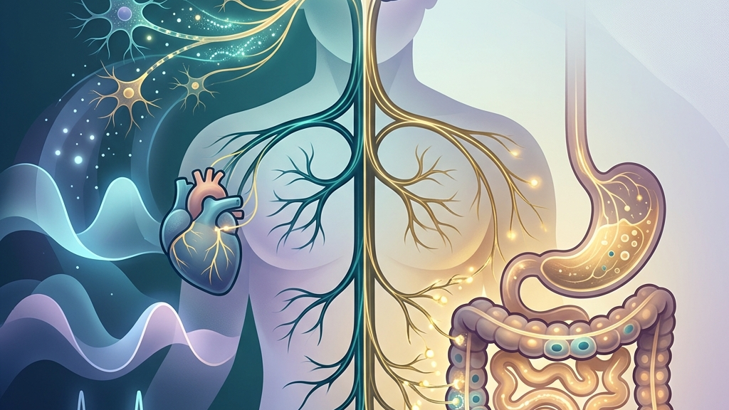
The vagus nerve serves as a primary conduit between the brainstem and major visceral organs, carrying signals that help coordinate rest and recovery with nutrient processing and motility. When this nerve maintains balanced tone, many people notice steadier transitions into deeper sleep stages alongside smoother digestion that feels less reactive after meals. Understanding these connections offers a clearer picture of why disruptions in one domain often appear alongside changes in the other. The nerve’s influence extends through everyday rhythms such as the gradual slowing of heart rate after a demanding workday or the gentle increase in intestinal contractions once a meal has settled, illustrating how autonomic balance supports both physical restoration and efficient processing of nutrients without requiring conscious effort.
Readers will explore the basic anatomy and signaling pathways of the vagus nerve, then examine its specific contributions to sleep continuity and gastrointestinal function. Evidence from physiological studies will illustrate measurable patterns, followed by gentle daily practices that may support vagal activity without promising outcomes. The discussion closes with guidance on when professional evaluation becomes important and answers to recurring questions. This progression allows a layered view of how sensory feedback from the body informs brainstem centers that in turn fine-tune organ function across the day and night.
How the Vagus Nerve Works
The vagus nerve emerges from the medulla oblongata as the tenth cranial nerve and extends long branches that reach the heart, lungs, esophagus, stomach, intestines, and other abdominal structures. It carries both afferent fibers that transmit sensory information upward to the brain and efferent fibers that deliver parasympathetic instructions downward to slow heart rate, stimulate digestive secretions, and promote smooth muscle relaxation. This bidirectional traffic forms a core part of the parasympathetic nervous system, often described as the “rest-and-digest” branch that counters sympathetic arousal. In practical terms, the efferent signals help the heart rate settle after climbing stairs while the afferent signals alert the brain when the stomach has filled, allowing adjustments in breathing depth or posture that further aid calm.
Within the gut-brain axis, vagal afferents monitor mechanical stretch, nutrient presence, and local inflammation, then relay these signals to brainstem nuclei that adjust autonomic output accordingly. Research on this pathway shows how vagal sensory neurons help integrate metabolic state with emotional and cognitive processing, creating a feedback loop that can influence both arousal levels before sleep and peristaltic rhythms after eating. Disruptions in this loop may appear as altered motility or fragmented rest, though individual patterns vary widely. For instance, after eating a fiber-rich lunch, stretch receptors along the intestinal wall send graded signals that can promote a slight drop in overall alertness, making an afternoon walk feel more restorative than stimulating.
Heart-rate variability serves as one accessible window into vagal tone because respiratory sinus arrhythmia—the natural speeding and slowing of the heartbeat with each breath—largely reflects vagal modulation of the sinoatrial node. Higher baseline variability often correlates with greater parasympathetic reserve, allowing quicker return to calm after mild stressors. Conversely, lower variability can accompany states of sustained sympathetic dominance, though many factors beyond the vagus contribute to these readings. Someone tracking morning readings might notice that a night of consistent sleep produces slightly larger swings between inhalation and exhalation beats, reflecting the cumulative effect of overnight recovery on autonomic flexibility.
How Vagal Activity Shapes Sleep Patterns
During the transition from wakefulness to light sleep, increased vagal outflow helps lower heart rate and respiratory rate, creating physiological conditions that favor entry into deeper non-REM stages. This parasympathetic shift reduces cortical arousal and supports the consolidation of slow-wave sleep, during which growth hormone release and tissue repair processes become more prominent. When vagal tone remains adequate, many individuals experience fewer micro-arousals and a more continuous progression through sleep cycles. A concrete example is the way a quiet evening routine of dim lighting and minimal screen time allows the natural lengthening of exhalations to reinforce this shift, making the first descent into deeper stages feel smoother rather than interrupted by residual mental activity.
Vagal afferents also participate in sensing metabolic signals that help time sleep pressure. As evening approaches, rising vagal activity may amplify the sense of satiety from daytime meals, indirectly supporting melatonin onset and the drop in core body temperature needed for sleep initiation. Studies examining sleep-disordered breathing note that vagal stimulation can influence upper-airway muscle tone and respiratory stability, suggesting a role in maintaining unobstructed airflow during vulnerable sleep phases. In daily life this might appear as the difference between falling asleep promptly after a balanced dinner versus lingering wakefulness after an unusually heavy or late meal that keeps digestive afferents more active.
Common observations linked to fluctuating vagal tone include difficulty settling at bedtime after high-stress days, when sympathetic drive remains elevated and heart-rate variability stays suppressed. Some people report that nights following large or late meals coincide with lighter sleep, possibly because digestive demands temporarily compete with vagal resources needed for full parasympathetic dominance. These patterns are not universal, yet they illustrate how the same nerve that quiets the heart also modulates the threshold for sleep maintenance. Another nuance appears when posture changes during the night; side-sleeping can gently stretch vagal branches near the esophagus, sometimes correlating with fewer position-related arousals compared with back-sleeping on particularly tense days.
Over longer periods, sustained low vagal tone may coincide with more frequent awakenings in the early morning hours, when cortisol naturally rises and sympathetic activity begins to predominate. Individuals who track subjective restfulness sometimes notice that periods of improved daytime calm correlate with fewer nighttime interruptions, consistent with the idea that baseline parasympathetic capacity influences sleep architecture without guaranteeing any specific outcome. Seasonal shifts offer further illustration: shorter winter days can reduce morning light exposure, subtly lowering average vagal responsiveness and making consistent wind-down routines more noticeable in their effect on continuity.
Vagal Signaling in the Digestive System
The vagus nerve supplies the majority of parasympathetic innervation to the stomach and proximal small intestine, releasing acetylcholine that stimulates gastric acid secretion, pancreatic enzyme release, and coordinated peristalsis. These motor commands facilitate mechanical breakdown and transit of food while also promoting local blood flow that supports mucosal integrity. Afferent fibers simultaneously report distension and nutrient composition back to the brainstem, allowing rapid adjustments in motility and secretion that match digestive load. After a meal containing both protein and complex carbohydrates, for example, the graded release of enzymes can feel like a steady rather than abrupt process, with hunger signals fading gradually instead of lingering.
Within the brain-gut axis, vagal pathways help integrate mechanical and chemical signals from the gut with higher-order regulation of appetite and satiety. When vagal tone is balanced, many people experience predictable hunger cues followed by comfortable fullness that does not linger into discomfort. Reduced vagal signaling, by contrast, can appear as delayed gastric emptying or irregular motility patterns that some describe as bloating or sluggish digestion after standard meals. Everyday nuance shows up when eating while distracted; the reduced afferent feedback can blunt the usual sense of satisfaction, leading to smaller second helpings or a longer period before the next natural hunger signal emerges.
Research on vagal sensory neurons highlights their role in detecting microbial metabolites and inflammatory mediators, which then influence brainstem autonomic centers and, indirectly, mood and energy regulation. This feedback may explain why periods of digestive unease sometimes coincide with changes in sleep depth or next-day alertness, though the direction of influence remains bidirectional and context-dependent. The same afferent traffic that informs the brain about gut state also participates in anti-inflammatory reflexes that dampen excessive immune activation along the intestinal wall. A practical illustration is the way a short post-meal walk can enhance these reflexes through mild mechanical stimulation along the digestive tract, sometimes resulting in more predictable bowel patterns the following morning.
Everyday experiences such as a sensation of throat tightness or a “knot” in the stomach during stress reflect vagal involvement in both visceral sensation and smooth-muscle tone. Because the nerve also innervates laryngeal and pharyngeal muscles, changes in vocal quality or swallowing ease can occasionally accompany shifts in overall vagal state, providing another observable clue to autonomic balance without serving as a diagnostic marker. Over weeks of noticing these patterns, some individuals link tighter vocal tone after rushed lunches with slightly heavier evening digestion, highlighting the interconnected daily loops the nerve helps regulate.
What the Research Shows
Clinical overviews describe the vagus nerve’s extensive distribution and its dual sensory-motor functions, underscoring its position as the longest cranial nerve and a major parasympathetic pathway. anatomical mapping of the vagus nerve illustrates how its branches reach both cardiac and gastrointestinal targets, providing the structural basis for coordinated regulation of heart rate and digestion.
Neuroanatomical reviews detail the nerve’s origin in the medulla and its passage through the jugular foramen before diverging into thoracic and abdominal branches. detailed cranial nerve mapping clarifies how vagal efferents modulate cardiac pacemaker activity while afferents convey visceral status, supporting the observed links between respiratory variability and gut motility.
Investigations into sleep-disordered breathing report associations between vagal stimulation parameters and improvements in sleep continuity measures. studies on vagus nerve stimulation and sleep quality examine respiratory and autonomic outcomes that may relate to vagal tone during nocturnal periods. Parallel work on heart-rate variability positions respiratory-linked fluctuations as a practical index of cardiac vagal tone. research on heart-rate variability and vagal tone outlines mechanisms by which parasympathetic modulation influences beat-to-beat intervals.
Additional studies explore the vagus nerve as a modulator within the brain-gut axis, showing how afferent traffic shapes central processing of gut-derived signals. work on the vagus nerve and brain-gut communication and investigations of vagal sensory neurons together describe pathways that link intestinal state with autonomic and behavioral adjustments relevant to both digestion and rest.
Practical Ways to Support Your Vagus Nerve
- Begin with slow, extended exhales that lengthen the out-breath relative to the in-breath; this pattern can increase respiratory sinus arrhythmia and thereby engage vagal pathways to the heart. Over several minutes the gradual change in intrathoracic pressure provides repeated gentle stimulation to baroreceptors, which in turn reinforce parasympathetic outflow without requiring special equipment.
- Practice gentle humming or soft gargling for a minute or two, which vibrates laryngeal muscles innervated by the vagus and may provide afferent feedback that supports parasympathetic tone. Choosing a comfortable pitch allows the vibration to travel along the nerve’s cervical branches, sometimes creating a noticeable sense of throat relaxation that carries into the next activity.
- Experiment with brief, tolerable cold exposure such as cool water on the face or a short cool shower at the end of a warm one, allowing gradual adaptation that some research associates with vagal activation. Starting with just the cheeks or forehead keeps the stimulus mild while still engaging trigeminal and vagal afferents that can influence heart-rate variability within minutes.
- Try paced breathing at roughly six breaths per minute for several minutes, a rate that often aligns with peak heart-rate variability and may reinforce vagal modulation of cardiac rhythm. Counting silently during both phases helps maintain consistency, and pairing the practice with a seated posture that keeps the spine neutral can enhance the mechanical effect on the chest wall.
- Incorporate light movement such as walking or gentle stretching after meals, which can stimulate vagal afferents through mechanical and postural changes along the digestive tract. A ten-minute stroll around the block after dinner, for example, combines mild gravitational shifts with rhythmic breathing that together support coordinated motility without intense effort.
- Maintain consistent morning light exposure within an hour of waking and protect a regular sleep window, both of which help align circadian cues that interact with vagal regulation of autonomic balance. Placing a chair near a window for the first cup of tea creates a repeatable anchor that supports the natural rise in morning parasympathetic tone.
When to Talk to a Professional
Sudden or severe changes in sleep continuity, such as repeated nighttime awakenings accompanied by breathing pauses, warrant medical assessment to rule out sleep-disordered breathing or other primary disorders. Likewise, persistent digestive symptoms including unexplained weight loss, blood in stool, or pain that interferes with daily function should prompt evaluation by a qualified clinician rather than reliance on self-directed practices.
Individuals experiencing chest pain, dizziness, or fainting should seek immediate care, as these can signal cardiac or autonomic issues that require prompt diagnosis. Any new or worsening symptoms that coincide with mood changes, swallowing difficulty, or voice alterations also merit professional attention to determine whether vagal or other neurological pathways are involved.
Common Questions
What exactly is vagal tone?
Vagal tone refers to the baseline level of parasympathetic activity carried by the vagus nerve, often inferred from heart-rate variability or physiological responses such as respiratory sinus arrhythmia. It is not a fixed trait but fluctuates with breathing, posture, and overall autonomic state. Daily variations can be observed when comparing readings taken after a restful versus a demanding morning, showing how recent activity shapes the current set point.
Can improving vagal tone guarantee better sleep or digestion?
While balanced vagal activity participates in the physiology of both sleep maintenance and gastrointestinal motility, many additional factors including circadian timing, diet composition, stress load, and underlying health conditions also shape outcomes. No single pathway determines results for everyone. Tracking personal patterns over several weeks often reveals interactions among these elements rather than isolated effects.
How long does it take to notice changes from daily practices?
Individual responses vary; some people report shifts in perceived calm or digestive comfort within days of consistent breathing or movement routines, while others observe more gradual adjustments over weeks. These timelines remain observational rather than prescriptive. Keeping a simple journal of sleep onset time or post-meal sensations can help identify personal trends without creating expectations.
Is there a difference between vagus nerve stimulation devices and natural practices?
Medical vagus nerve stimulation involves implanted or external devices delivering electrical pulses under clinical supervision, whereas everyday practices such as breathing or cold exposure represent non-invasive attempts to engage similar pathways. The two approaches differ substantially in mechanism, intensity, and evidence base. Device-based methods are typically reserved for specific clinical indications, while natural practices remain accessible entry points for general autonomic awareness.
Does age affect vagal function?
Research indicates that average heart-rate variability tends to decline with advancing age, yet lifestyle, fitness level, and health status influence this trend more than chronological age alone. Individual assessment remains necessary rather than assumptions based on years lived. Comparing one’s own readings across seasons or after changes in routine often provides more relevant information than population averages.
Have a question?
Have a question about something specific? Send us a message.
Visit VagusSkool.com/contact — we'll try to get back to you within 24 hours.