The Vagus Nerve as a Central Regulator of Sleep, Stress Recovery, and Digestion
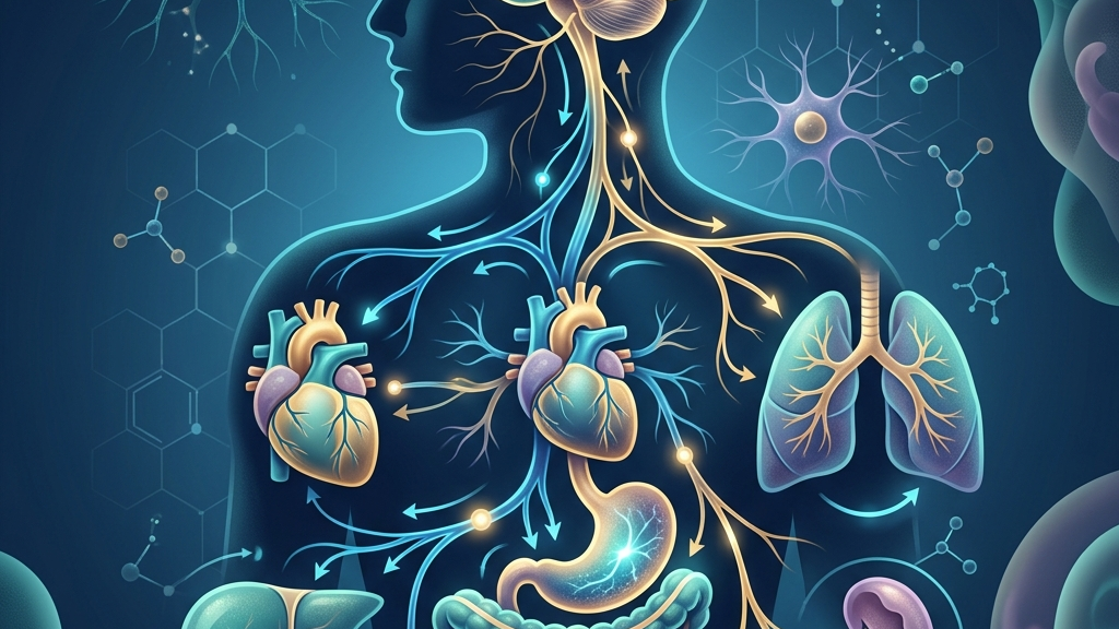
The vagus nerve serves as a primary communication pathway between the brain and many internal organs, influencing how the body transitions between states of activity and rest. Because it carries both sensory and motor signals, it helps coordinate processes that unfold over hours or days, such as the depth of nighttime rest, the speed of return to calm after pressure, and the efficiency of nutrient processing. Understanding these connections offers a clearer picture of why disruptions in one area often appear alongside changes in the others. For example, someone who experiences frequent work deadlines may notice that elevated evening heart rate coincides with both slower digestion after dinner and more fragmented sleep later, illustrating how a single neural highway can link seemingly separate daily rhythms. The nerve’s mixed fiber composition allows it to transmit precise data about blood pressure fluctuations in the aortic arch or stretch in the stomach wall, enabling the brainstem to fine-tune output accordingly rather than issuing blanket commands.
Readers will encounter the basic architecture of the nerve, followed by detailed examinations of its contributions to each of the three topics. Mechanisms are presented alongside observations commonly reported in daily life, always framed as possibilities rather than certainties. Later sections review published findings and outline simple, low-demand practices that some individuals incorporate into routines. The goal is to supply context for informed conversations with clinicians when needed. In practice this means recognizing that a person who feels persistently “wired” after evening exercise might explore whether reduced vagal rebound is one contributing factor among many, including caffeine timing or room temperature, before drawing conclusions.
How the Vagus Nerve Works
The vagus nerve emerges from the medulla oblongata and extends through the neck, chest, and abdomen, branching extensively to reach the heart, lungs, esophagus, stomach, and intestines. As the principal parasympathetic outflow, it promotes conservation of energy by slowing heart rate, stimulating digestive secretions, and supporting restorative functions during periods of safety. Its long course allows it to sample conditions in multiple organ systems and relay that information upward, creating a continuous feedback loop that helps the brain adjust autonomic tone moment to moment. Consider the everyday sequence after finishing a large meal: gastric stretch receptors activate vagal afferents that signal satiety to the nucleus tractus solitarius, which in turn increases vagal efferent traffic to slow the heart slightly and enhance pancreatic enzyme release, illustrating how one nerve can orchestrate both local gut responses and systemic cardiovascular adjustments within minutes.
Within the gut-brain axis, roughly 80 percent of vagal fibers transmit signals from the viscera to the brainstem rather than the reverse. These afferent fibers detect mechanical stretch, chemical composition of luminal contents, and local immune activity, then influence brainstem nuclei that modulate mood, arousal, and inflammation. The result is a bidirectional channel through which intestinal states can shape central nervous system activity and vice versa. A concrete illustration occurs when bacterial metabolites such as short-chain fatty acids bind to receptors on enteroendocrine cells; the resulting vagal firing can subtly shift brainstem noradrenergic tone, which some people experience as a gentle post-meal dip in alertness rather than drowsiness caused solely by blood glucose changes.
Heart-rate variability, particularly the high-frequency component tied to respiration, serves as one accessible window into vagal cardiac regulation. When vagal outflow increases, the interval between heartbeats lengthens on exhalation; when it decreases, the interval shortens. This dynamic range reflects the nerve’s capacity to apply a “brake” that can be released quickly when demands rise and reapplied when safety returns. In daily terms, someone sitting quietly at a desk may observe that their smart-watch HRV reading rises during a relaxed phone call with a friend, then drops sharply when an unexpected work email arrives, demonstrating the nerve’s rapid recalibration without any conscious effort.
The Vagus Nerve and Restorative Sleep
During the shift from wakefulness to non-REM sleep, vagal activity tends to rise, contributing to the slowing of heart rate and respiratory rhythm that characterize deeper sleep stages. This increase supports the reduction of sympathetic tone and may facilitate the release of hormones and neurotransmitters involved in tissue repair and memory consolidation. Because the nerve also innervates upper-airway muscles, its tone can influence airway patency, an effect explored in studies of sleep-disordered breathing. A person who practices a wind-down routine of reading in low light may notice that their resting heart rate drops more steadily after 10 p.m., potentially reflecting greater vagal engagement that coincides with fewer micro-arousals once they fall asleep.
People sometimes notice that evenings marked by lower physiological arousal are followed by shorter sleep latency and fewer nighttime awakenings. Conversely, when vagal withdrawal persists into the night, the same individuals may describe lighter sleep or a sense of unrefreshing rest even after adequate time in bed. These patterns are not universal and can be modulated by many factors, including age, medications, and concurrent health conditions. For instance, a parent who checks work messages late may experience prolonged sympathetic dominance that delays the usual evening rise in vagal tone, leading to more stage-one sleep and a greater likelihood of waking to small household sounds.
Respiratory sinus arrhythmia, an index of vagal influence on the heart, often becomes more pronounced during slow-wave sleep. This amplification suggests that the nerve participates in the autonomic stabilization required for sustained deep sleep. When this stabilization is less robust, transitions between sleep stages may become more fragmented, though the precise contribution of vagal tone versus other systems remains under investigation. Athletes sometimes report that nights following intense late-day training show reduced RSA amplitude, which can correspond to more frequent shifts between deep and light stages even when total sleep time appears adequate on a tracker.
Chronic low-grade inflammation can dampen vagal signaling, and some researchers have proposed that this damping may indirectly affect sleep continuity by altering brainstem arousal thresholds. The relationship is complex and likely involves additional pathways, yet it illustrates how systemic states can feed back onto nightly rest through vagal routes. Someone managing a persistent low-grade sinus issue might observe that weeks of mild inflammation coincide with both slightly elevated morning resting heart rate and subjectively lighter sleep, highlighting one possible indirect route without implying direct causation.
Vagal Tone in Recovering from Stress
After an acute stressor ends, the vagus nerve helps re-engage parasympathetic dominance, lowering heart rate and blood pressure while restoring digestive motility that may have been suppressed. This recovery phase depends on the nerve’s ability to inhibit sympathetic outflow and to dampen inflammatory cytokine production via the cholinergic anti-inflammatory pathway. When vagal tone is higher at baseline, the return to equilibrium after challenge tends to occur more smoothly. A teacher finishing a challenging parent conference may feel their shoulders drop and stomach rumble return within twenty minutes once the meeting concludes, consistent with vagal reactivation that permits both cardiovascular deceleration and renewed gut motility.
Many people report that sensations of chest tightness or abdominal unease linger after demanding events; these experiences can coincide with periods of reduced vagal modulation. As vagal activity rebounds, such sensations often diminish, although the timeline varies widely. The speed of this rebound is one element reflected in heart-rate-variability measurements taken during recovery windows. An office worker who steps outside for five minutes of quiet breathing after a tense call may notice the tightness easing more quickly than on days when they immediately open the next email thread.
Prolonged or repeated stress can produce lasting shifts in vagal regulation, including lower resting variability and slower return of parasympathetic activity after challenge. These shifts are not deterministic and can be influenced by sleep quality, physical activity patterns, and social context. Research continues to examine how sustained changes in vagal tone relate to subjective feelings of resilience or depletion. Caregivers who juggle multiple roles over months sometimes describe a gradual sense that ordinary stressors take longer to “shake off,” a pattern that can parallel measurable reductions in baseline HRV without any single cause being identifiable.
The nerve’s connections to laryngeal and pharyngeal muscles also allow vocalization and breath patterns to influence vagal afferents, providing a route through which deliberate changes in breathing or sound production may support the transition out of heightened alert states. Such effects remain modest and context-dependent rather than guaranteed. Singing along to music in the car after a difficult day offers one informal example where rhythmic vocalization and extended exhalation occur together, sometimes followed by a subjective easing that aligns with the same physiological window used in more structured breathing exercises.
Vagal Pathways in Digestive Regulation
The vagus nerve supplies parasympathetic innervation to the esophagus, stomach, small intestine, and proximal colon, promoting peristalsis, gastric acid secretion, and pancreatic enzyme release under resting conditions. Afferent fibers from these organs convey information about nutrient density, mechanical distension, and local inflammation back to the nucleus tractus solitarius, shaping subsequent autonomic and behavioral responses such as satiety signaling and food-seeking. Eating a balanced lunch while seated away from screens allows vagal afferents to register gradual gastric filling and transmit graded satiety signals, often resulting in a natural stopping point before over-distension occurs.
Individuals sometimes observe that meals eaten in a calm setting are followed by more comfortable digestion, whereas hurried or tense eating coincides with bloating or irregular motility. These differences may partly reflect vagal withdrawal during sympathetic activation and subsequent re-engagement once the meal is finished. The magnitude of such effects differs across people and situations. A commuter who eats breakfast while driving may later experience mid-morning sluggishness or mild reflux that is less common on days when the same meal is consumed while seated at a table with attention on the food.
Within the gut-brain axis, vagal sensory neurons respond to microbial metabolites and enteroendocrine signals, transmitting data that can influence central appetite circuits and mood-related pathways. This traffic is not one-way; descending vagal signals can alter intestinal barrier function and immune cell activity, illustrating a loop that links emotional and digestive states over longer timescales. Someone who notices afternoon mood dips after several days of irregular, rushed meals may be experiencing one expression of this extended feedback, though many other factors such as sleep debt and hydration also contribute.
Disruptions in vagal gastrointestinal traffic, whether from surgical resection, neuropathy, or sustained high sympathetic tone, can manifest as altered gastric emptying or changes in bowel regularity. Such outcomes depend on many variables, and any persistent digestive change warrants clinical evaluation rather than self-interpretation through a vagal lens alone. Patients recovering from certain abdominal procedures sometimes track gradual return of normal motility patterns over weeks, underscoring that vagal contributions interact with local tissue healing and medication effects.
What the Research Shows
Investigations using both animal models and human recordings have documented that vagal stimulation can increase the proportion of slow-wave sleep and reduce the frequency of respiratory events during sleep in selected populations, as summarized in research on vagus nerve stimulation, sleep-disordered breathing, and sleep quality. These findings remain preliminary for broad application and require replication across larger cohorts. Human studies often employ non-invasive transcutaneous approaches at the ear or neck, allowing researchers to observe changes in sleep architecture without implanted devices, yet the magnitude of benefit varies with stimulation parameters and individual baseline autonomic profiles.
Heart-rate-variability studies consistently link higher resting vagal indices to faster cardiovascular recovery after laboratory stressors, detailed in work on heart rate variability and cardiac vagal tone. The same measures also correlate, though modestly, with self-reported calm in daily life, underscoring that variability captures only one dimension of autonomic flexibility. Longitudinal observations in working adults show that days with higher morning HRV often precede evenings with quicker post-work decompression, although lifestyle confounders such as physical activity and social support remain difficult to isolate completely.
Reviews of the brain-gut axis emphasize that vagal afferents constitute a major route for microbial and nutrient signals to reach brainstem and forebrain structures, outlined in analysis of the vagus nerve as modulator of the brain-gut axis. Complementary anatomical work maps the specific sensory neuron populations responsible for these signals, presented in studies of vagal sensory neurons and gut-brain signaling. Optogenetic techniques in rodents have clarified that distinct vagal neuron subtypes convey stretch versus nutrient information, providing a cellular basis for why identical meals can produce different satiety timelines depending on prior autonomic state.
Foundational anatomical descriptions from clinical sources confirm the nerve’s extensive thoracic and abdominal distribution and its mixed sensory-motor composition, available in Cleveland Clinic material on vagus nerve function and location and NIH StatPearls neuroanatomy of cranial nerve 10. Together these sources illustrate both the breadth of vagal influence and the limits of current interventional evidence. Cadaveric tracing and modern diffusion imaging together reveal extensive branching that explains why irritation or compression at one cervical level can produce sensations referred to thoracic or abdominal structures.
Practical Ways to Support Your Vagus Nerve
- Slow, extended exhales performed for several minutes can transiently increase heart-rate variability by lengthening the expiratory phase during which vagal cardiac inhibition is strongest.
- Humming or gentle gargling engages laryngeal muscles innervated by the vagus, potentially augmenting afferent traffic that supports parasympathetic rebound after periods of concentration.
- Brief, tolerable cold exposure such as cool water on the face or hands may activate vagal afferents through trigeminal and cervical pathways, though individual tolerance varies considerably.
- Paced breathing at roughly six breaths per minute aligns with the frequency at which baroreflex-vagal coupling is most efficient, offering a low-effort method some people use before sleep.
- Light movement such as walking after meals can promote gastrointestinal motility partly through vagal recruitment while avoiding the intensity that might sustain sympathetic dominance.
- Consistent morning light exposure combined with stable sleep timing helps anchor circadian rhythms that in turn influence nightly vagal dominance during sleep.
When to Talk to a Professional
Sudden or severe changes in heart rhythm, persistent difficulty swallowing, unexplained weight loss, or intense abdominal pain should prompt medical assessment regardless of any vagal considerations. Likewise, sleep disturbances accompanied by witnessed breathing pauses, excessive daytime sleepiness, or mood changes that interfere with daily function merit evaluation by a qualified clinician. These symptoms can arise from multiple causes, and only a professional can determine appropriate next steps. Early consultation allows for comprehensive testing that distinguishes vagal contributions from structural, endocrine, or medication-related factors.
Common Questions
Can vagal tone be measured at home?
Consumer devices that estimate heart-rate variability provide indirect approximations of cardiac vagal modulation, yet these readings are influenced by breathing pattern, posture, and recent activity; they do not replace clinical assessment. Morning orthostatic measurements taken under consistent conditions can reveal day-to-day trends, but single readings remain noisy indicators best interpreted alongside sleep logs and symptom diaries.
Does everyone experience the same effects from breathing practices?
Responses vary with age, fitness level, medications, and concurrent conditions; some individuals notice clearer shifts in perceived calm while others observe minimal change, underscoring the value of personalized guidance. Older adults or those taking beta-blockers may show smaller acute HRV increases from the same breathing protocol compared with younger, unmedicated volunteers.
Are there risks to stimulating the vagus nerve?
Most simple practices such as slow breathing carry low risk for healthy adults, but people with certain heart conditions, epilepsy, or recent neck surgery should consult a physician before attempting more intensive techniques. Individuals with implanted cardiac devices in particular require medical clearance before exploring electrical or strong mechanical stimulation methods.
How long does it take to notice changes?
Acute shifts in heart-rate variability can appear within minutes of a breathing exercise, whereas longer-term patterns tied to sleep or digestion typically unfold over weeks and depend on many intersecting factors. Consistent tracking over a month often reveals whether a given practice aligns with improved subjective energy or comfort more reliably than isolated sessions.
The vagus nerve offers one integrative lens on the interplay among nightly rest, return to baseline after challenge, and gastrointestinal function. While the mechanisms are increasingly well mapped, individual outcomes remain variable and context-dependent. Tracking personal patterns and seeking professional input for concerning symptoms together provide the most grounded approach to supporting these interconnected systems.
Have a question?
Have a question about something specific? Send us a message.
Visit VagusSkool.com/contact — we'll try to get back to you within 24 hours.