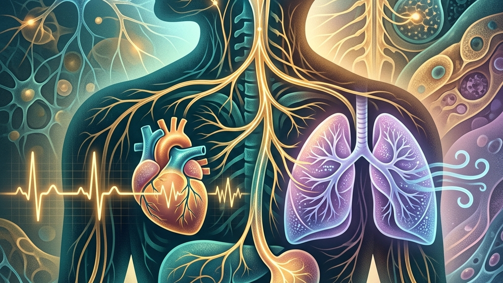
The Vagus Nerve as a Bridge Between Stress Recovery, Resting Heart Rhythms, and Breath
The vagus nerve occupies a central position in how the body moves between states of alertness and restoration. Its extensive reach allows it to influence heart rhythm, digestive signaling, and respiratory control in ways that shape daily resilience. Understanding these connections offers a clearer picture of why certain patterns of breathing or heart-rate changes often accompany shifts in perceived stress levels. The nerve functions as a bidirectional highway, transmitting both outgoing commands that slow physiological processes and incoming reports from organs that update the brainstem about current internal conditions. This constant exchange supports the transition from high-alert sympathetic dominance after a challenge back to a calmer baseline, where energy can be directed toward maintenance rather than immediate action. Everyday observations, such as noticing a calmer stomach after a walk or a steadier pulse during quiet reading, often reflect this underlying traffic without the individual realizing the specific neural route involved.
Readers will encounter the basic anatomy and signaling routes of the nerve, followed by focused examinations of its involvement in stress recovery, resting heart-rate regulation, and breath. Later sections review published observations, outline accessible daily practices, note situations that call for professional input, and address recurring questions. The goal is to map physiological mechanisms without implying guaranteed outcomes for any individual. Each section builds on the previous one by adding layers of mechanical detail and real-world context, illustrating how small, repeated inputs can accumulate into noticeable shifts in daily function. Attention remains on the interplay between the nerve and other systems rather than isolated effects, providing a framework for recognizing patterns that appear across different times of day or activity levels.
How the vagus nerve works
The vagus nerve emerges from the medulla oblongata and travels through the neck, chest, and abdomen, carrying both sensory and motor fibers. As the primary parasympathetic outflow, it promotes conservation of energy by slowing heart rate, supporting digestion, and modulating inflammatory responses. Its bidirectional traffic means signals travel from peripheral organs back to the brainstem, creating continuous feedback loops that help maintain internal balance. Motor fibers release acetylcholine at target organs to reduce contraction force in the heart or increase motility in the intestines, while sensory fibers return data on stretch, pressure, and chemical composition. This loop operates largely below conscious awareness yet influences decisions such as when to pause eating or how long to linger in a relaxed posture after a meal.
One well-described route is the gut-brain axis, where vagal afferents relay information about nutrient status, microbial activity, and mechanical stretch in the intestines to brainstem nuclei. This information can influence mood-related circuits and autonomic tone. Research on this pathway highlights how changes in visceral state may register as shifts in overall arousal or calm without requiring conscious awareness. For instance, after consuming a large meal, distension signals travel via the nerve and can prompt a subtle reduction in alertness, encouraging rest rather than continued activity. Conversely, an empty stomach may send signals that heighten vigilance, preparing the body for foraging or movement. These visceral updates integrate with other inputs such as light exposure and sleep history, producing a composite autonomic tone that varies from morning to evening.
Heart-rate variability serves as an accessible window into vagal function. Higher variability at rest often reflects stronger parasympathetic modulation, allowing the heart to respond flexibly to changing demands. Lower variability can appear when sympathetic drive predominates or when vagal outflow is reduced. These patterns arise from the interplay between the vagus and sympathetic nerves at the sinoatrial node, where acetylcholine release lengthens the interval between beats. In daily life this appears as a slightly irregular pulse during relaxed reading or conversation, versus a more metronomic rhythm during focused work or after caffeine intake. The variability itself is not random; it follows predictable respiratory and postural influences that can be observed by simply feeling the pulse at the wrist while breathing normally.
Vagal Pathways Supporting Recovery from Stress
After an acute stressor, the vagus nerve contributes to the return toward baseline by re-engaging parasympathetic control over cardiovascular and gastrointestinal systems. This re-engagement can dampen lingering sympathetic activation, allowing heart rate and blood pressure to settle. The speed of this shift varies with existing vagal capacity, prior sleep, and concurrent breathing patterns. Recovery is rarely instantaneous; instead it unfolds over minutes as afferent signals from baroreceptors and lung stretch receptors accumulate and gradually inhibit sympathetic outflow. Individuals may notice the transition when swallowing becomes easier or when abdominal sounds return after a period of quiet.
Mechanistically, vagal afferents detect changes in blood pressure and lung volume, feeding information to the nucleus tractus solitarius. From there, inhibitory projections reach sympathetic premotor neurons, reducing catecholamine release. At the same time, vagal efferents to the heart increase acetylcholine, lengthening diastolic intervals and lowering oxygen demand. These coordinated actions create a physiological environment more conducive to tissue repair and cognitive reappraisal. In practice this might look like the gradual easing of shoulder tension while sitting quietly after a difficult meeting, accompanied by a measurable drop in heart rate over the following five to ten minutes.
Many people notice that after periods of elevated tension, sensations such as a slower pulse, easier swallowing, or renewed digestive activity coincide with feeling less keyed up. These subjective shifts often track with measurable increases in heart-rate variability during recovery windows. The nerve does not eliminate the memory of stress, yet its activity can shorten the duration of physiological after-effects. The process resembles releasing a brake that had been partially applied; once the brake pressure increases again, other systems follow rather than leading the change.
Chronic or repeated stressors may gradually reduce vagal responsiveness if recovery intervals remain insufficient. In such cases, the baseline tone available for subsequent challenges can decline, lengthening the time needed to regain equilibrium. This dynamic underscores why consistent opportunities for physiological downregulation matter over longer timescales. For example, scheduling brief pauses between back-to-back video calls allows vagal afferents to register lower demand and begin restoring tone before the next demand arrives.
Resting Heart Rate and the Vagal Brake
At rest, the vagus nerve exerts a tonic braking effect on the sinoatrial node, keeping heart rate below the intrinsic pacemaker rate of roughly 100 beats per minute. This continuous influence produces the slower resting rates commonly observed in trained athletes or individuals with high vagal tone. Withdrawal of the brake during activity allows rapid acceleration without requiring immediate sympathetic recruitment. The brake operates through ongoing acetylcholine release that hyperpolarizes pacemaker cells, extending the time before the next spontaneous depolarization occurs.
The strength of this braking action can be inferred from respiratory sinus arrhythmia, the natural fluctuation in heart rate that occurs with each breath. Inhalation briefly reduces vagal tone while exhalation restores it, creating beat-to-beat variability. Stronger fluctuations generally indicate greater resting vagal influence and a wider dynamic range for responding to internal or external demands. This can be observed by counting pulse intervals during quiet sitting: intervals lengthen slightly on exhale and shorten on inhale, producing a gentle oscillation rather than a fixed rhythm.
Observations in both clinical and non-clinical groups show that resting heart rates in the mid-50s to low-70s often accompany higher baseline variability, whereas rates consistently above 80 may reflect lower vagal contribution or elevated sympathetic tone. These associations are correlational; many factors, including fitness level, age, and medication use, also shape resting values. Posture provides an everyday modulator: lying supine typically increases vagal braking compared with standing, which is why resting heart-rate measurements are standardized to a seated or reclined position.
Over time, repeated activation of the vagal brake during quiet periods may support more stable blood-pressure regulation and lower myocardial workload. Conversely, sustained suppression of vagal outflow can leave the heart under predominantly sympathetic control even at rest, narrowing the margin for further acceleration when needed. Tracking personal resting trends alongside breathing habits can therefore illuminate shifts in autonomic balance. A person who records morning pulse rates over several weeks may notice that days with longer evening wind-down periods show modestly lower values the following morning.
Breathing Rhythms and Vagus Nerve Activity
Respiration directly modulates vagal outflow through mechanical and neural routes. Lung stretch receptors and thoracic pressure changes send afferent signals that influence brainstem nuclei governing cardiac vagal neurons. Slow, extended exhales tend to augment vagal tone, lengthening heart-rate intervals, while rapid or shallow patterns can reduce it. The mechanical effect arises because prolonged exhalation increases intrathoracic pressure slightly, which in turn enhances baroreceptor firing and subsequent vagal activation.
The anatomical proximity of vagal branches to the larynx and pharynx adds another layer. Rhythmic vocal cord vibration or controlled throat engagement can stimulate these fibers, producing measurable changes in heart-rate variability. This connection explains why certain sound-based or breath-focused practices often coincide with subjective calm and objective shifts in autonomic markers. Even simple activities such as reading aloud or singing softly can engage the same pathways through repeated laryngeal movement.
People commonly report that deliberate slowing of the breath, particularly with longer exhales, brings a sense of settling within a few minutes. These reports align with documented increases in high-frequency heart-rate variability during paced breathing at rates near six breaths per minute. The effect is not uniform; individual differences in lung capacity, posture, and current arousal level influence the magnitude of change. Someone with a history of shallow breathing may require several practice sessions before the same exhale duration produces a comparable shift.
Over repeated sessions, consistent breathing practice may raise the average level of vagal tone available during daily activities. This does not eliminate stressors but can enlarge the physiological window in which recovery processes operate. Respiratory patterns therefore serve as both a readout and a modifiable input to vagal regulation. Noticing whether breath naturally lengthens after a meal or shortens during focused work provides ongoing feedback about current autonomic state.
What the research shows
Studies examining cranial nerve anatomy confirm that the vagus supplies parasympathetic innervation to thoracic and abdominal viscera while carrying extensive sensory information back to the brainstem, as detailed in Neuroanatomy, Cranial Nerve 10 (Vagus Nerve). This architecture underpins its role in moment-to-moment adjustments of heart rate and gut motility. Additional mapping studies have traced how individual fascicles within the nerve bundle serve distinct organ targets, allowing selective influence rather than blanket effects across all viscera.
Investigations into heart-rate variability demonstrate that higher resting vagal tone correlates with greater beat-to-beat flexibility, described in Heart Rate Variability and Cardiac Vagal Tone. These patterns appear sensitive to both acute challenges and longer-term lifestyle factors. Longitudinal observations further indicate that variability can fluctuate within a single day according to posture, recent food intake, and emotional context, underscoring the value of repeated measurements rather than single snapshots.
Research on the gut-brain axis shows vagal sensory neurons convey microbial and nutrient signals that can influence central autonomic networks, outlined in Vagus Nerve as Modulator of the Brain–Gut Axis and Vagal Sensory Neurons and Gut–Brain Signaling. Such findings help explain why gastrointestinal comfort often tracks with overall relaxation. Experiments that alter gut microbiota in animal models have shown corresponding changes in vagal firing rates and downstream behavioral markers, suggesting a feedback route that operates continuously in humans as well.
Additional work on sleep and breathing reports associations between vagal stimulation parameters and improved sleep architecture in certain populations, presented in Vagus Nerve Stimulation, Sleep-Disordered Breathing & Sleep Quality. Cleveland Clinic summaries further note the nerve’s broad distribution and its involvement in multiple regulatory loops, available at Vagus Nerve: Function, Location & Conditions. These summaries emphasize that the nerve’s extensive branching creates numerous points at which everyday behaviors can intersect with its signaling.
Practical ways to support your vagus nerve
- Slow extended exhales performed for several minutes while seated can lengthen heart-rate intervals by briefly enhancing vagal outflow to the heart.
- Humming or gentle gargling creates rhythmic vibration near vagal branches in the throat, which some individuals find accompanies a drop in perceived tension.
- Brief, tolerable cold exposure such as cool water on the face may activate vagal afferents and produce a measurable shift in heart-rate variability within minutes.
- Paced breathing at approximately six breaths per minute aligns respiratory and cardiac rhythms, often increasing high-frequency variability during the session.
- Light movement such as walking after meals can stimulate vagal traffic through both mechanical gut signals and modest cardiovascular demand.
- Consistent morning light exposure combined with stable sleep timing supports circadian alignment that indirectly favors higher average vagal tone across the day.
When to talk to a professional
Sudden or persistent changes in heart rhythm, unexplained fainting, severe swallowing difficulty, or rapidly worsening digestive symptoms warrant prompt medical evaluation. These signs may reflect issues unrelated to vagal tone and require assessment by a qualified clinician. The same principle applies to any new symptom cluster that appears abruptly or intensifies over days rather than weeks.
Individuals experiencing intense anxiety that interferes with daily function, chronic insomnia, or chest pain should also seek professional guidance rather than relying solely on self-directed breathing or lifestyle adjustments. Early consultation helps rule out underlying conditions that need targeted management. Tracking symptom patterns over time, including time of day and relation to meals or activity, can provide useful information for the clinician during an evaluation.
Common questions
How quickly can breathing changes affect heart-rate variability?
Observable shifts in variability can appear within one to two minutes of adopting slower, longer exhales, though the size of the change differs across people and depends on current autonomic state. The rapidity arises because respiratory afferents reach brainstem nuclei within a single breath cycle, allowing almost immediate adjustment of cardiac vagal neurons.
Does higher resting heart-rate variability always indicate better health?
Higher variability at rest generally reflects stronger vagal modulation, yet it remains one marker among many; overall health involves additional systems and cannot be reduced to a single measure. Factors such as medication, recent illness, and genetic variation also contribute to recorded values, so interpretation benefits from context rather than isolated numbers.
Can gut discomfort influence vagal signaling?
Yes, vagal afferents carry information from the digestive tract that can modulate brainstem autonomic centers, sometimes registering as shifts in overall arousal or calm. Mechanical distension or changes in luminal contents provide ongoing input that the nerve integrates with other visceral and environmental signals.
Are there age-related changes in vagal tone?
Vagal influence tends to decline gradually with advancing age, yet regular movement, breathing practice, and sleep routines may help maintain a portion of that capacity. Cross-sectional studies show wide individual variation at every decade, indicating that lifestyle patterns can offset some of the average age-related trend.
Is vagus nerve stimulation only available through devices?
Medical-grade stimulation requires clinical devices, while everyday activities such as paced breathing or vocalization can engage vagal pathways without specialized equipment. The distinction lies in intensity and precision: device-based approaches deliver controlled electrical pulses, whereas behavioral methods rely on natural afferent routes that are accessible at any time.
The vagus nerve offers a tangible route for understanding how stress recovery, resting heart rhythms, and breath remain interconnected. Attention to these relationships can inform steadier daily patterns while underscoring the value of professional care when symptoms exceed ordinary variation.
Have a question?
Have a question about something specific? Send us a message.
Visit VagusSkool.com/contact — we'll try to get back to you within 24 hours.