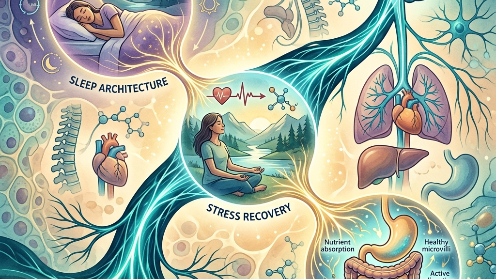
The Vagus Nerve as a Bridge Between Sleep Architecture, Stress Recovery, and Digestive Function
How the Vagus Nerve Works
The vagus nerve, designated cranial nerve X, originates in the medulla oblongata and extends long branches that reach the heart, lungs, esophagus, stomach, small intestine, and portions of the colon. Roughly eighty percent of its fibers are afferent, conveying sensory data from visceral organs back to the brainstem, while the remaining efferent fibers carry parasympathetic instructions outward. This bidirectional traffic allows the nerve to participate in rapid adjustments of heart rate, respiratory rhythm, and gastrointestinal motility without requiring conscious direction. In its parasympathetic capacity the vagus nerve supplies the primary brake on sympathetic arousal. When vagal outflow increases, acetylcholine release at cardiac ganglia lengthens the interval between heartbeats, raising heart-rate variability. The same neurotransmitter acts on enteric neurons to promote peristalsis and glandular secretion while dampening local inflammatory signaling through the cholinergic anti-inflammatory pathway. These actions occur continuously, yet their amplitude fluctuates with respiration, posture, and metabolic state. For instance, after a large meal the efferent signals intensify to coordinate gastric mixing and intestinal transit, whereas during upright posture or mild exertion the balance tilts toward modest withdrawal to permit appropriate cardiovascular support without overshooting into sustained acceleration. The gut-brain axis illustrates the nerve’s integrative role most clearly. Vagal afferents in the intestinal mucosa detect mechanical stretch, nutrient composition, and microbial metabolites, then relay that information to the nucleus tractus solitarius. From there, ascending projections influence hypothalamic and limbic structures involved in satiety, mood, and sleep pressure. Reciprocal descending signals modulate vagal efferent tone, closing a loop that couples digestive status with central autonomic balance. Research on heart-rate variability and cardiac vagal tone underscores how measurable shifts in this loop correspond to changes in both emotional regulation and visceral function. A concrete illustration appears when someone consumes a fiber-rich meal: short-chain fatty acids generated by fermentation stimulate enteroendocrine release of glucagon-like peptide-1, which in turn activates nearby vagal endings and ultimately dampens hypothalamic appetite signaling while simultaneously nudging cardiac intervals toward greater variability.Vagal Tone and the Architecture of Sleep
During non-rapid-eye-movement sleep the parasympathetic branch becomes dominant, and vagal activity contributes to the progressive slowing of heart rate and respiratory rate that characterizes deeper stages. Afferent signals from pulmonary stretch receptors and baroreceptors travel via the vagus to reinforce brainstem circuits that sustain slow-wave activity. When vagal tone is robust, the transition into slow-wave sleep tends to occur more smoothly and the duration of consolidated deep sleep lengthens, supporting the clearance of metabolic by-products from neural tissue. Respiratory sinus arrhythmia, a direct expression of vagal modulation, reaches its highest amplitude during slow-wave sleep. This oscillation reflects moment-to-moment adjustments in cardiac vagal outflow synchronized with breathing. Studies that record both polysomnography and continuous electrocardiography show that individuals with higher baseline respiratory sinus arrhythmia often exhibit fewer micro-arousals and more stable oxygen saturation across the night. Conversely, reduced vagal modulation appears alongside increased sleep fragmentation, although the direction of causality remains under investigation. In practical terms, someone who finishes a demanding evening shift may notice that an extended period of quiet sitting before bed allows the arrhythmia amplitude to build, whereas immediate exposure to bright screens can blunt the same oscillation by sustaining low-level sympathetic drive. People commonly notice that evenings marked by lower physiological arousal are followed by quicker sleep onset and fewer nocturnal awakenings. The subjective sense of “restorative” sleep frequently coincides with mornings in which heart-rate variability measured upon waking remains elevated, suggesting that nocturnal vagal recovery carried forward. Light exposure timing, meal composition, and evening wind-down routines can each shift the balance of sympathetic and parasympathetic input, thereby altering the substrate on which vagal regulation operates during sleep. Nuance arises when considering that not every high-variability night produces identical subjective refreshment; factors such as prior sleep debt or ambient temperature interact with vagal tone to determine how efficiently slow-wave activity clears adenosine and other sleep-promoting substances.Vagal Contributions to Recovery from Stress
Stress recovery depends on the timely re-engagement of vagal efferents after sympathetic activation has subsided. The vagus nerve supplies inhibitory projections to the sinoatrial node that counteract residual tachycardia and support the return of heart-rate variability to pre-stress levels. This “vagal brake” operates through both tonic and phasic mechanisms; tonic tone sets the resting capacity for deceleration, while phasic bursts allow rapid adjustments when environmental demands change. When vagal pathways remain partially withdrawn after a stressor, recovery of emotional and physiological equilibrium slows. Prolonged reduction in cardiac vagal tone is associated with sustained low-grade inflammation and altered baroreflex sensitivity, both of which can feed forward into further autonomic imbalance. Conversely, moments of increased vagal outflow—such as those elicited by extended exhalation or vocalization—accelerate the dissipation of sympathetic after-effects and restore a wider dynamic range to heart-rate variability. An everyday example occurs after an unexpected work deadline: the initial surge in heart rate and muscle tension may linger for minutes or hours unless interrupted by a deliberate slowing of breath, which recruits vagal fibers to shorten the recovery window and reduce the likelihood of carry-over effects into subsequent tasks. Individuals often describe a subjective shift from vigilance to calm once vagal tone reasserts itself. This shift may manifest as easier access to prefrontal regulatory circuits, reduced muscle tension, and a gradual normalization of digestive sensations that had been suppressed during sympathetic dominance. Because vagal afferents also convey information about peripheral inflammatory status, the resolution of stress-related arousal frequently coincides with a perceptible quieting of interoceptive signals that previously registered as unease. The nuance here lies in recognizing that complete return to baseline rarely occurs in a single step; instead, oscillatory re-engagement of the brake allows incremental restoration while the system monitors ongoing environmental cues.The Vagus Nerve in Digestive Regulation
Within the gastrointestinal tract the vagus nerve supplies both motor and secretory control. Efferent fibers stimulate gastric motility, pancreatic enzyme release, and bile flow while coordinating the migrating motor complex that clears residual contents between meals. Afferent fibers, densely distributed in the mucosal and submucosal layers, monitor luminal contents and mechanical distension, transmitting satiety and discomfort signals that shape subsequent ingestive behavior. The same afferent traffic participates in the gut-brain axis modulation of central autonomic tone. Metabolites produced by the microbiota can activate vagal endings either directly or through enteroendocrine cells, influencing brainstem nuclei that in turn adjust vagal outflow to the gut. This closed loop allows digestive activity to modulate its own neural regulation and, by extension, to influence sleep propensity and stress responsiveness. Consider the difference between eating a balanced lunch in a quiet environment versus hurriedly consuming the same food while checking messages: the former permits fuller vagal engagement that times gastric emptying appropriately and signals satiety before overconsumption occurs, whereas the latter often suppresses motility enough to produce later sensations of fullness or irregular transit. People commonly observe that meals taken in a calm setting are followed by more complete digestion and less postprandial fatigue. When vagal tone is adequate, gastric emptying proceeds at a measured pace, reducing the likelihood of reflux or bloating. Conversely, periods of sustained sympathetic activation can suppress vagally mediated motility, leading to sensations of heaviness or irregular bowel patterns that resolve once autonomic balance is restored. These visceral experiences feed back into the same afferent pathways, reinforcing or attenuating the original autonomic state. Additional nuance appears in the timing of the migrating motor complex, which requires several hours of relative fasting and low sympathetic tone to sweep indigestible material forward; habitual late-night snacking can truncate these cycles and subtly alter next-day appetite signaling via the same vagal routes.What the Research Shows
Investigations using both animal models and human cohorts have mapped the anatomical substrates and functional consequences of vagal signaling. descriptions of vagus nerve anatomy and parasympathetic distribution establish the structural foundation for its widespread influence. Complementary neuroanatomical work in detailed cranial nerve mapping clarifies the medullary origins and visceral projections that enable coordinated cardiorespiratory and gastrointestinal control. Human studies examining sleep have linked vagal metrics to objective sleep parameters. research on vagus nerve stimulation and sleep-disordered breathing reports associations between enhanced vagal activity and improved sleep continuity in selected populations. Parallel work on autonomic markers demonstrates that higher resting heart-rate variability, an index of cardiac vagal tone, correlates with more efficient transitions between sleep stages. Evidence concerning stress recovery centers on the dynamic properties of vagal withdrawal and re-engagement. analyses of heart-rate variability and cardiac vagal tone document how rapid restoration of variability after laboratory stressors predicts lower subsequent emotional reactivity. In the gastrointestinal domain, reviews of the vagus nerve as modulator of the brain–gut axis synthesize findings that afferent vagal traffic shapes both peripheral inflammation and central autonomic set-points. Additional mechanistic studies in work on vagal sensory neurons and gut–brain signaling detail the cellular pathways by which luminal signals reach the brainstem. Collectively these sources indicate that vagal function operates at the intersection of the three domains rather than within isolated silos. Effect sizes vary across studies, and many rely on correlational designs; nevertheless, the consistency of directional findings supports continued exploration of vagal pathways as a common physiological substrate. The research also underscores that laboratory measures such as respiratory sinus arrhythmia amplitude or baroreflex gain provide only snapshots, and real-world translation benefits from repeated ambulatory recordings that capture how daily posture changes, meal timing, and social interactions modulate the same metrics.Practical Ways to Support Your Vagus Nerve
- Slow, extended exhales performed for several minutes while seated can lengthen the interval between heartbeats and temporarily elevate heart-rate variability by increasing vagal outflow to the sinoatrial node.
- Humming or gentle gargling engages laryngeal branches of the vagus and may produce a brief rise in parasympathetic tone through mechanical stimulation of vagal afferents in the throat.
- Brief, tolerable cold exposure such as cool water on the face or a short cool shower activates thermoregulatory reflexes that recruit vagal pathways to stabilize cardiovascular parameters after the initial sympathetic response.
- Paced breathing at approximately six breaths per minute aligns respiratory and cardiac rhythms, amplifying respiratory sinus arrhythmia and thereby providing a measurable window of heightened vagal modulation.
- Light movement such as walking after meals supports vagally mediated gastric motility by combining mild postural change with rhythmic afferent input from the lower limbs.
- Consistent morning light exposure together with a stable sleep schedule helps entrain circadian oscillators that in turn influence the daily rhythm of vagal tone across sleep and wakefulness.
When to Talk to a Professional
Symptoms that appear suddenly or intensify rapidly merit medical evaluation regardless of any autonomic considerations. These include severe chest pain, pronounced difficulty swallowing, persistent vomiting, unexplained weight loss, or breathing disturbances during sleep that cause daytime somnolence. Changes in bowel habits accompanied by bleeding, fever, or significant pain also warrant prompt assessment. A clinician can determine whether further testing of cardiac, neurological, or gastrointestinal function is indicated and can place any observed autonomic patterns within a broader diagnostic context. The value of such evaluation lies in distinguishing transient fluctuations in vagal tone from underlying structural or inflammatory conditions that require targeted intervention.Common Questions
Does stimulating the vagus nerve guarantee better sleep?
Research suggests associations between higher vagal tone and more stable sleep architecture, yet individual responses vary and no single practice ensures a specific sleep outcome. Factors such as accumulated sleep pressure, ambient noise, and concurrent medication use can override acute changes in respiratory sinus arrhythmia, so practices function best as supportive elements within a broader sleep-hygiene framework rather than isolated levers.
Can vagal pathways influence both digestion and mood simultaneously?
Because vagal afferents convey information from the gut to brainstem nuclei that project to limbic and hypothalamic regions, changes in digestive signaling can coincide with shifts in affective and autonomic state. This convergence occurs through shared second-order neurons in the nucleus tractus solitarius, allowing a single meal’s nutrient profile or microbial activity to register both as altered gastric comfort and as subtle adjustments in perceived energy or emotional tone.
How quickly might someone notice changes after trying breathing practices?
Acute shifts in heart-rate variability can appear within minutes of slow breathing, while longer-term patterns of stress recovery or digestive comfort typically unfold over days to weeks of consistent use. The rapid component reflects immediate acetylcholine release at cardiac ganglia, whereas sustained adaptations involve gradual recalibration of baroreflex sensitivity and possibly modest changes in vagal fiber excitability with repeated recruitment.
Is there a risk of overstimulating the vagus nerve?
Excessive or abrupt stimulation can produce transient lightheadedness or bradycardia in susceptible individuals; therefore any new practice should begin gently and be discontinued if uncomfortable symptoms arise. Susceptibility often relates to baseline autonomic tone or concurrent medications that already lengthen cardiac intervals, underscoring the importance of starting with short durations and monitoring subjective response.
Do medications affect vagal function?
Certain medications can alter heart-rate variability or gastrointestinal motility through direct or indirect effects on autonomic signaling; discussion with a prescriber clarifies individual implications. Beta-blockers, for example, may blunt measurable respiratory sinus arrhythmia without eliminating underlying vagal traffic, while prokinetic agents can enhance motility partly by amplifying efferent vagal effects on enteric neurons. The interplay among sleep, stress recovery, and digestion through vagal pathways illustrates how a single neural structure participates in multiple regulatory loops. Attention to the conditions that support balanced vagal activity—respiratory rhythm, circadian timing, and manageable daily demands—offers a coherent perspective for exploring these connections further while remaining attentive to professional guidance when symptoms require it.
Have a question?
Have a question about something specific? Send us a message.
Visit VagusSkool.com/contact — we'll try to get back to you within 24 hours.