The Vagus Nerve and Stress Recovery: How Parasympathetic Pathways Help Restore Balance
Stress is an unavoidable part of life, yet the speed and completeness with which the body returns to equilibrium after a stressful event vary widely from person to person. At the center of this recovery process sits the vagus nerve, the longest cranial nerve and primary conductor of the parasympathetic nervous system. Understanding its anatomy and signaling patterns offers a window into why some individuals rebound quickly while others remain in a heightened state long after the stressor has passed.
This article examines the vagus nerve’s contributions to stress recovery through its influence on heart rhythm, sleep architecture, digestive signaling, and vocal-motor pathways. Readers will encounter the underlying physiology that links vagal activity to measurable signs such as heart-rate variability, explore how everyday patterns like sleep and vocal use intersect with vagal tone, and review the current state of evidence drawn from neuroanatomical and clinical studies. The goal is to provide a clear, mechanism-focused foundation rather than prescriptive advice.
By tracing these connections, the discussion highlights both the nerve’s reach and its limitations, underscoring that while vagal function is modifiable through daily behaviors, individual responses differ and professional evaluation remains essential when symptoms persist or intensify.
How the vagus nerve works
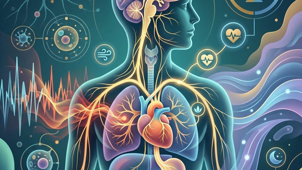
The vagus nerve originates in the medulla oblongata and extends through the neck, thorax, and abdomen, carrying both efferent fibers that slow heart rate and promote digestion and afferent fibers that relay sensory information from the viscera back to the brain. As the principal parasympathetic outflow, it counterbalances sympathetic activation by releasing acetylcholine at target organs, which lowers heart rate, reduces inflammatory signaling, and supports gastrointestinal motility. This bidirectional traffic forms a continuous feedback loop that helps the nervous system gauge internal conditions and adjust autonomic tone accordingly.
Within the gut-brain axis, vagal afferents transmit mechanical and chemical signals from the intestinal mucosa to brainstem nuclei, influencing mood-regulating centers and hypothalamic-pituitary-adrenal axis activity. Research on these pathways shows that vagal stimulation can modulate cytokine production and alter the excitability of brain regions involved in threat appraisal. The same nerve also contributes to heart-rate variability, the beat-to-beat fluctuation that reflects the dynamic interplay between sympathetic acceleration and parasympathetic braking; higher variability generally corresponds to greater vagal influence and more flexible stress responses.
Because the vagus nerve interfaces with multiple organ systems simultaneously, its activity level shapes how quickly physiological arousal subsides once a stressor ends. When vagal tone is robust, heart rate decelerates smoothly, digestion resumes, and inflammatory markers remain contained. When tone is reduced, sympathetic dominance lingers, producing prolonged elevations in heart rate, shallower breathing, and disrupted sleep continuity. These patterns illustrate why vagal function is often discussed in the context of recovery rather than acute stress itself.
Sleep and Vagal Tone
During non-REM sleep stages, parasympathetic dominance increases and vagal cardio-motor neurons become more active, producing the characteristic slowing of heart rate and rise in heart-rate variability observed in healthy sleep cycles. This nocturnal shift allows the cardiovascular system to rest and supports the clearance of metabolic byproducts accumulated during waking hours. When stress disrupts sleep architecture, the expected nocturnal rise in vagal activity is blunted, leaving heart-rate variability lower than usual and extending the time required for daytime recovery.
Vagal afferents from the gut also participate in sleep regulation by conveying satiety and inflammatory signals that influence brainstem sleep centers. Elevated evening cytokines, for instance, can fragment sleep and further suppress vagal outflow, creating a cycle in which poor sleep and reduced vagal tone reinforce each other. Over successive nights, this cycle may manifest as morning fatigue, elevated resting heart rate, and difficulty shifting out of a vigilant state even in safe environments.
People commonly notice that nights of fragmented sleep are followed by a sense of emotional reactivity or physical tension that takes longer to dissipate. Conversely, nights marked by deeper, more continuous sleep often coincide with a subjective feeling of steadiness the following day. These observations align with the physiological role of vagal reactivation during sleep as one component of overnight autonomic recalibration.
Because sleep and vagal tone are mutually reinforcing, any factor that protects sleep continuity indirectly supports the parasympathetic recovery processes that unfold across the night. The relationship is bidirectional: sustained stress can erode sleep quality, while eroded sleep quality can diminish vagal capacity for the next day’s demands.
Resting Heart Rate and the Vagal Brake
The vagus nerve exerts a tonic inhibitory influence on the sinoatrial node, often described as the vagal brake. At rest, this brake keeps heart rate below the intrinsic pacemaker rate and permits rapid adjustments when metabolic demand changes. During stress recovery, re-engagement of the brake produces a prompt deceleration of heart rate and an increase in heart-rate variability as parasympathetic outflow resumes dominance. When vagal tone is low, the brake remains partially disengaged, resulting in a persistently elevated resting heart rate and reduced variability even after the original stressor has subsided.
Heart-rate variability metrics such as root mean square of successive differences and high-frequency power serve as indirect markers of vagal cardio-motor activity. Studies of these indices demonstrate that individuals with higher resting variability tend to exhibit faster return to baseline cardiovascular parameters after laboratory stressors. Lower variability, by contrast, correlates with slower recovery and prolonged sympathetic activation, which can be perceived as lingering physical arousal or difficulty settling into relaxed states.
Over time, a chronically elevated resting heart rate may reflect cumulative reductions in vagal braking capacity. People sometimes describe this as a feeling of being “revved up” without an obvious cause, or as needing longer periods of quiet before heart rate settles. These subjective reports correspond to the measurable loss of parasympathetic modulation that occurs when vagal efferent traffic is diminished.
Restoration of the vagal brake is therefore one physiological target in discussions of stress recovery. Because heart rate is continuously modulated by both autonomic branches, any improvement in vagal tone can be observed as a gradual lowering of resting heart rate and a widening of its variability across the day, provided other factors such as fitness and medication remain constant.
Voice, Throat, and the Vagus Nerve
Branchial motor fibers of the vagus nerve innervate the intrinsic muscles of the larynx and pharynx, controlling vocal fold tension, swallowing, and the gag reflex. These same fibers participate in the neural control of prosody and vocal pitch, which are subtly altered during states of autonomic arousal. When vagal tone is adequate, laryngeal muscle activity supports a relaxed vocal quality; when tone decreases, subtle increases in muscle tension can produce a higher-pitched or strained voice that listeners may perceive as tense or fatigued.
Afferent fibers traveling through the superior and recurrent laryngeal nerves provide sensory feedback from the throat to the brainstem, contributing to the regulation of breathing patterns and the coordination of swallowing with respiration. This sensory traffic helps maintain the balance between airway protection and efficient gas exchange. Stress-related changes in breathing depth and rhythm can therefore influence vagal afferent load, creating another route by which psychological strain registers in vagal signaling.
Individuals often notice that periods of heightened stress coincide with throat tightness, a tendency to clear the throat more frequently, or a voice that tires more quickly during conversation. These sensations reflect both the motor and sensory roles of the vagus and can persist until autonomic balance is restored. Because the laryngeal muscles are under direct vagal control, practices that engage gentle vocalization or swallowing can transiently increase vagal afferent traffic and support a return toward parasympathetic dominance.
The throat thus serves as both an output pathway for vagal motor commands and an input pathway for sensory information that helps calibrate overall autonomic state. Disruptions in this region may therefore signal and sustain alterations in vagal tone that affect broader stress-recovery processes.
What the research shows
Neuroanatomical mapping confirms that the vagus nerve constitutes the primary parasympathetic supply to thoracic and abdominal viscera, establishing its central role in autonomic regulation. Neuroanatomy, Cranial Nerve 10 (Vagus Nerve) details the nerve’s medullary origin and bilateral distribution, providing the structural basis for its influence on heart rate and gastrointestinal function during recovery from stress.
Clinical investigations of heart-rate variability demonstrate that higher vagally mediated variability predicts faster cardiovascular recovery after acute stressors. Heart Rate Variability and Cardiac Vagal Tone reviews the physiological mechanisms linking vagal outflow to beat-to-beat fluctuations and discusses how reduced variability may index impaired parasympathetic regulation. Parallel work on the gut-brain axis shows that vagal afferents convey inflammatory and metabolic signals capable of modulating central stress circuitry. Vagus Nerve as Modulator of the Brain–Gut Axis and Vagal Sensory Neurons and Gut–Brain Signaling describe these pathways and their relevance to autonomic balance.
Evidence also connects vagal activity to sleep quality. Vagus Nerve Stimulation, Sleep-Disordered Breathing & Sleep Quality examines how altered vagal signaling may contribute to sleep fragmentation, while Vagus Nerve: Function, Location & Conditions summarizes clinical conditions in which vagal dysfunction is implicated. Together these sources indicate that vagal tone participates in multiple overlapping systems that support or hinder return to baseline after stress, although the magnitude of effect varies across individuals and contexts.
Practical ways to support your vagus nerve
- Slow, extended exhales that lengthen the out-breath relative to the in-breath can increase vagal cardio-motor activity and are often among the simplest entry points for noticing shifts in heart-rate variability.
- Humming or gentle gargling engages laryngeal muscles innervated by the vagus and may transiently raise vagal afferent traffic through the throat.
- Brief, tolerable cold exposure such as cool water on the face can stimulate vagal sensory endings and is frequently reported to produce a calming sensation shortly afterward.
- Paced breathing at approximately six breaths per minute aligns with resonance frequencies that amplify heart-rate oscillations driven by the vagus.
- Light movement such as walking after meals supports gastrointestinal motility partly through vagal efferent pathways and may aid the postprandial return toward parasympathetic dominance.
- Consistent morning light exposure combined with stable sleep timing helps align circadian rhythms that in turn influence nocturnal vagal reactivation during sleep.
When to talk to a professional
Persistent resting heart rates above 100 beats per minute, repeated episodes of dizziness or fainting, or sudden changes in swallowing or voice that do not resolve warrant medical evaluation to rule out structural or neurological issues affecting the vagus nerve. Severe or rapidly worsening digestive symptoms, unexplained weight loss, or chest pain should also prompt consultation, as these may indicate conditions beyond normal variations in autonomic tone.
When sleep disturbance is accompanied by profound daytime fatigue, mood changes, or breathing pauses observed by others, a sleep study or cardiopulmonary assessment may be appropriate. Individuals with known cardiovascular disease, diabetes, or autoimmune conditions should discuss any new autonomic symptoms with their clinician, because vagal pathways intersect with multiple medical systems.
Professional guidance ensures that any observed changes in vagal-related markers are interpreted within the full clinical picture rather than in isolation.
Common questions
How long does it take to notice changes in vagal tone?
Acute shifts in heart-rate variability can appear within minutes of a breathing or postural change, yet sustained alterations in baseline tone generally develop over weeks of consistent practice and depend on factors such as age, fitness, and concurrent health conditions.
Can vagal tone be measured at home?
Consumer devices that estimate heart-rate variability provide approximate trends but lack the precision of clinical electrocardiography; interpretation should remain cautious and not replace professional assessment.
Is there an upper limit to beneficial vagal activity?
Excessive parasympathetic dominance can produce bradycardia or syncope in susceptible individuals, illustrating that autonomic balance rather than maximal vagal tone is the relevant target for most people.
Do medications affect vagal function?
Certain drugs, including beta-blockers and anticholinergics, directly alter vagal effects on the heart; any consideration of interactions belongs in a conversation with a prescribing clinician.
Does age change vagal recovery capacity?
Vagal tone tends to decline gradually with advancing age, yet the rate of decline varies and can be influenced by physical activity patterns and overall health status.
Stress recovery is not a single switch but an ensemble of physiological processes in which the vagus nerve plays a recurring, integrative part. By supporting the conditions under which vagal signaling can operate—stable sleep, rhythmic breathing, and gentle sensory engagement—many people find that their return to baseline after stress becomes more reliable, even if the nerve itself remains only one element within a larger adaptive system. When symptoms exceed ordinary variation, professional evaluation provides the necessary context for understanding what is occurring and what steps may be appropriate next.
Have a question?
Have a question about something specific? Send us a message.
Visit VagusSkool.com/contact — we'll try to get back to you within 24 hours.