The Vagus Nerve and Its Interwoven Roles in Sleep, Stress Recovery, and Digestion
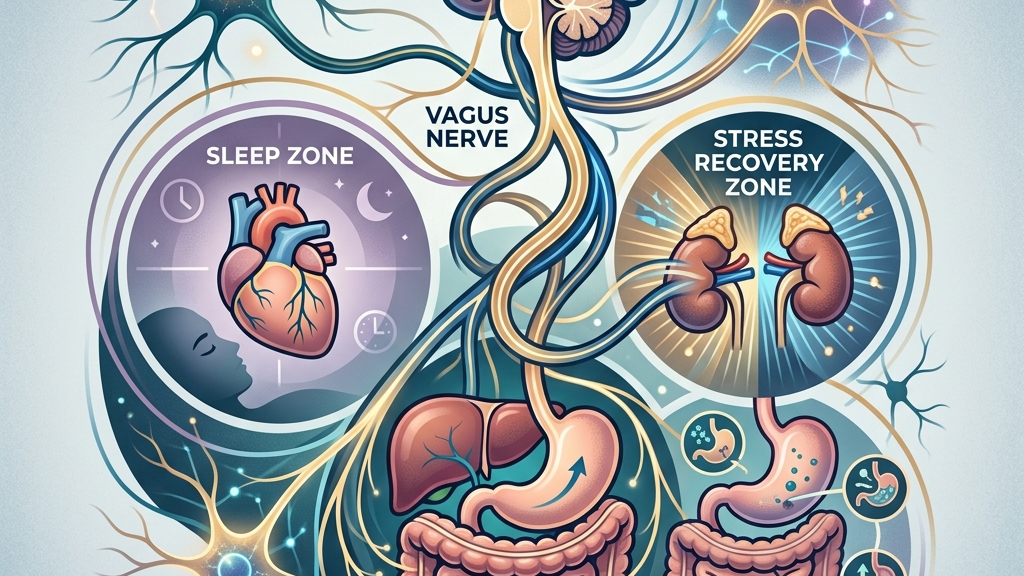
The vagus nerve forms one of the body’s primary communication highways between the brain and many internal organs. Its activity influences how readily the system shifts into rest-and-repair states, how efficiently the heart and lungs coordinate during both activity and stillness, and how the digestive tract processes nutrients without excessive tension. Understanding these connections offers a clearer picture of why sleep quality, emotional recovery after pressure, and everyday digestive comfort often move together rather than in isolation. The nerve achieves this coordination through a dense network of myelinated and unmyelinated fibers that release acetylcholine at target organs, dampening excitability in cardiac pacemaker cells, relaxing bronchial smooth muscle, and enhancing peristaltic waves along the gastrointestinal tract. In daily life this shows up when someone finishes a large meal and feels their pulse slow while intestinal sounds become audible, or when a person sits quietly after an argument and notices their breathing deepen without conscious effort.
This article examines the nerve’s basic anatomy and signaling patterns, then explores its contributions to each of the three areas in turn. Mechanisms are described in physiological terms, and common subjective experiences are noted without implying that any single practice will produce a particular outcome. Later sections review selected research findings, outline accessible daily habits that some people explore, and clarify when professional evaluation is appropriate. Throughout, the emphasis remains on how bidirectional signaling integrates information from multiple organ systems so that changes in one domain, such as an evening of screen time that delays melatonin onset, can ripple into nighttime heart-rate patterns and next-day appetite regulation.
How the vagus nerve works
The vagus nerve is the tenth cranial nerve and the longest of the parasympathetic nerves. It originates in the brainstem and travels through the neck, chest, and abdomen, sending and receiving signals to the heart, lungs, esophagus, stomach, intestines, and other structures. Its parasympathetic fibers promote a baseline state of reduced heart rate, slower breathing, and increased digestive motility when the body is not under immediate threat. This “rest-and-digest” bias is not an on-off switch but a continuous modulation that competes with sympathetic arousal. At the cellular level, preganglionic neurons in the dorsal motor nucleus and nucleus ambiguus release acetylcholine onto postganglionic neurons located near each target organ; those postganglionic fibers then liberate acetylcholine again, binding muscarinic receptors on cardiac myocytes to lengthen the interval between beats or on enteric neurons to increase the frequency of slow-wave contractions. In everyday terms, this mechanism operates when a person reclines after lunch and feels their heart rate drop five to ten beats per minute while stomach churning becomes noticeable, illustrating the same pathway that also quiets bronchial tone so breathing feels less effortful.
Information travels in both directions. Sensory fibers from the gut, heart, and lungs carry data about stretch, inflammation, and chemical conditions back to the brainstem, while motor fibers adjust organ function in response. This bidirectional traffic constitutes the core of the gut-brain axis. When vagal sensory traffic is steady and varied, the brain receives an ongoing map of internal conditions that supports appropriate adjustments in arousal and digestion. For instance, after eating a fiber-rich salad, mechanoreceptors in the stomach wall signal distension while chemoreceptors detect short-chain fatty acids produced by microbiota; these signals ascend via the vagus to the nucleus tractus solitarius, which then modulates descending autonomic output so that subsequent meals feel satisfying rather than urgent. Conversely, during an afternoon of back-to-back meetings, reduced afferent variety from a clenched abdomen can leave the brainstem with less precise data, making it slightly harder to downshift into relaxed breathing once the meetings end.
Heart-rate variability (HRV) provides one measurable window into vagal influence. Higher variability at rest often reflects stronger parasympathetic modulation of the sinoatrial node, sometimes described as a “vagal brake.” Lower resting variability can occur when sympathetic tone predominates or when vagal pathways are less active. Researchers therefore use HRV as an indirect index of vagal tone, though many other factors, including breathing rate and fitness level, also shape the reading. In practice, someone who tracks nightly HRV on a wearable may notice that readings rise on evenings after a thirty-minute walk and fall after late caffeine intake; the difference arises because rhythmic locomotion stimulates pulmonary stretch receptors whose afferents travel in the vagus, briefly amplifying respiratory sinus arrhythmia and leaving a modest residual increase in beat-to-beat interval variation once the person lies down.
How Vagal Tone Supports Restorative Sleep
During the transition into sleep, parasympathetic dominance typically increases. Vagal outflow helps lower heart rate and blood pressure while promoting slower, deeper breathing. This shift reduces cortical arousal and supports the progression from light to deeper non-REM stages. When vagal signaling is robust, many people notice that their heart rate settles more quickly once they lie down and that breathing becomes less effortful in the first hour after sleep onset. Mechanistically, the baroreflex arc is strengthened: each heartbeat stretches carotid and aortic baroreceptors whose afferents also travel partly via the vagus, triggering further acetylcholine release that lengthens the subsequent diastolic interval. An everyday illustration is the difference between falling asleep after an evening of light reading versus after an intense workout; in the former case the baroreflex remains responsive, so heart rate drifts downward within minutes, whereas post-exercise sympathetic metabolites can keep the same reflex partially inhibited for longer.
Throughout the night the vagus nerve continues to influence autonomic balance. Brief arousals or shifts in body position trigger small sympathetic bursts; intact vagal pathways help return the system to parasympathetic dominance afterward. Research on sleep-disordered breathing suggests that vagal sensory feedback from the airways and lungs participates in stabilizing respiratory rhythm during sleep, though the precise contribution varies with individual anatomy and health status. Consider a person who wakes briefly because a pet jumps on the bed; within seconds the vagus conveys updated lung-volume information that allows the brainstem to resume the slower respiratory pattern characteristic of stage N2, preventing a cascade into full wakefulness. When this feedback loop is less efficient, the same minor disturbance may produce a longer period of elevated heart rate before sleep resumes.
Subjectively, people sometimes describe nights when they fall asleep more readily after periods of calm daytime activity. Others observe that evenings spent in sustained mental or physical activation are followed by more fragmented rest. These patterns align with the idea that daytime vagal engagement can leave a residual bias toward parasympathetic tone at bedtime, though many additional factors, including light exposure and room temperature, also affect sleep architecture. A concrete example is the office worker who takes two short walks during lunch and again mid-afternoon; the accumulated afferent traffic from limb muscles and thorax tends to support a smoother descent into slow-wave sleep, whereas the same person who remains seated for eight hours may experience more frequent micro-arousals even if total sleep time appears unchanged on a tracker.
Disrupted vagal signaling does not act alone. Inflammatory signals, pain, or medications can alter both vagal traffic and sleep continuity. Therefore, changes in sleep quality are best viewed as one possible downstream effect within a larger network rather than a direct result of vagal function in isolation. For example, a mild upper-respiratory infection increases local cytokine release that can blunt vagal afferent sensitivity from the lungs, making it harder for the brainstem to maintain stable breathing patterns overnight; the resulting sleep fragmentation is thus multifactorial even though vagal pathways are involved.
The Vagus Nerve and Recovery from Stress
Stress recovery depends in part on the speed and completeness with which parasympathetic activity reasserts itself after a sympathetic surge. The vagus nerve supplies a rapid pathway for this reassertion by slowing heart rate and reducing vascular tone once the immediate demand has passed. Individuals with higher baseline vagal tone often show quicker return of heart rate to resting levels after laboratory stressors, a pattern sometimes called vagal rebound. In daily terms this appears when someone receives an unexpected work email at 4 p.m.; if vagal pathways are responsive, heart rate may peak and then decline within five minutes once the initial reaction passes, whereas lower tone can leave the same person with an elevated pulse for twenty minutes or longer while they continue typing.
Respiratory sinus arrhythmia, the natural fluctuation in heart rate that accompanies breathing, is largely vagally mediated. Slow, rhythmic breathing can temporarily amplify this fluctuation and, over repeated practice, may support a modest increase in resting HRV for some people. The effect is not uniform; age, fitness, and concurrent health conditions all modulate the response. Still, the physiological principle remains that extended exhalation tends to increase vagal outflow to the heart. A person sitting in traffic can test this by consciously lengthening each out-breath to six seconds while keeping inhalation at four; within a minute the pulse typically becomes more variable, illustrating how the same mechanism operates during a brief pause before entering a stressful meeting.
Many people report that after a demanding day they feel “wired but tired,” a state in which mental fatigue coexists with physical activation. This experience can correspond to incomplete vagal reactivation, leaving sympathetic tone relatively elevated even though the original challenge has ended. Gentle movement, such as walking, sometimes facilitates the shift by engaging vagal afferents from the limbs and thorax without adding new sympathetic load. The contrast is noticeable when someone chooses a slow stroll around the block rather than remaining seated at a desk; the rhythmic limb feedback helps the nucleus tractus solitarius recalibrate autonomic set-points so that subsequent relaxation feels more complete.
Chronic or repeated stressors can down-regulate vagal responsiveness over time, a change reflected in lower HRV readings in some populations. This adaptation is not permanent in every case; reductions in daily demand or the introduction of consistent recovery practices may allow partial restoration, though individual trajectories differ and are influenced by many variables beyond vagal pathways alone. Longitudinal observations show that people who move from high-pressure jobs to roles with clearer boundaries sometimes record gradual HRV increases over months, yet the same shift may be slower or absent when sleep debt or inflammatory conditions persist.
Vagal Pathways in Digestive Function and Comfort
The vagus nerve supplies the majority of parasympathetic innervation to the stomach and small intestine. Motor fibers stimulate gastric motility and acid secretion while sensory fibers relay information about distension, nutrient composition, and inflammation back to the brainstem. This loop helps coordinate the timing of digestive phases and contributes to the sensation of satiety once a meal has begun. After a balanced plate of protein, vegetables, and whole grains, for instance, progressive gastric filling activates stretch receptors whose vagal signals reach the brainstem within minutes, prompting both a gradual slowing of eating rate and a later release of satiety peptides that reinforce the sense of having had enough.
When vagal tone is adequate, many individuals experience steady gastric emptying and regular peristalsis without pronounced urgency or sluggishness. Conversely, periods of high sympathetic activation can reduce vagal outflow, slowing motility and altering secretion patterns. The result may be a temporary sense of fullness, bloating, or irregular bowel habits that resolves once autonomic balance is restored. A common illustration occurs during a high-stakes presentation: sympathetic dominance diverts resources away from digestion, so lunch consumed an hour earlier may still feel heavy two hours later; once the presentation ends and vagal tone rebounds, gastric emptying resumes and the sensation of fullness fades.
Vagal sensory neurons also participate in signaling from gut microbiota metabolites and enteroendocrine hormones. These signals influence brainstem nuclei that in turn modulate appetite, mood, and further autonomic output. The complexity of this traffic means that digestive comfort reflects an interplay among neural, hormonal, and microbial factors rather than vagal activity in isolation. Short-chain fatty acids produced by fermentation of dietary fiber, for example, bind receptors on enteroendocrine cells that release GLP-1 and PYY; vagal afferents carry this information centrally, contributing to both the decision to stop eating and a subtle shift toward calmer mood that many notice after a fiber-rich meal.
Common observations include changes in digestion during travel or after abrupt schedule shifts. In such situations, disrupted sleep and elevated sympathetic tone often coincide with altered appetite or bowel patterns. While these experiences are familiar, they arise from multiple converging influences, and any single factor such as vagal tone should not be viewed as the sole determinant. Jet lag that advances the circadian clock by six hours can simultaneously suppress melatonin, elevate evening cortisol, and reduce the amplitude of vagal rhythms that normally support nocturnal peristalsis, producing the familiar combination of constipation and restless sleep until the new schedule stabilizes.
What the research shows
Studies using HRV as a proxy for cardiac vagal tone have documented associations between higher resting variability and faster cardiovascular recovery after laboratory stress tasks, as summarized in Heart Rate Variability and Cardiac Vagal Tone. These findings do not establish causation but illustrate how vagal modulation can be quantified and tracked over time. Additional work has examined how acute changes in posture or breathing rate alter the same metric within a single session, underscoring that HRV reflects moment-to-moment integration of respiratory, baroreflex, and central influences.
Research on sleep-disordered breathing has explored vagus nerve stimulation as one experimental avenue for improving respiratory stability during sleep, detailed in Vagus Nerve Stimulation, Sleep-Disordered Breathing & Sleep Quality. Results remain preliminary and vary by stimulation parameters and patient characteristics. Parallel observational studies have noted that individuals with higher baseline HRV tend to show fewer oxygen-desaturation events, though the direction of influence and the role of confounding variables such as body weight require further clarification.
Investigations into the gut-brain axis emphasize the vagus nerve’s role in transmitting microbial and nutrient signals that influence central autonomic networks, as reviewed in Vagus Nerve as Modulator of the Brain–Gut Axis and Vagal Sensory Neurons and Gut–Brain Signaling. These pathways help explain why changes in gut environment can coincide with shifts in arousal and digestion, though translation to everyday experience requires further study. Animal models have demonstrated that selective vagotomy abolishes certain behavioral responses to microbial metabolites, highlighting the nerve’s necessity in those specific circuits while leaving open questions about redundancy with hormonal routes in humans.
Anatomical descriptions from sources such as Vagus Nerve: Function, Location & Conditions and Neuroanatomy, Cranial Nerve 10 (Vagus Nerve) provide the foundational map of fiber distribution that underpins the functional observations above. These references detail the precise branching pattern through the jugular foramen, the cardiac and pulmonary plexuses, and the anterior and posterior gastric branches, supplying the structural basis for the physiological mechanisms described throughout this article.
Practical ways to support your vagus nerve
- Slow, extended exhales during quiet sitting or lying down can temporarily increase vagal outflow to the heart by lengthening the expiratory phase of the respiratory cycle.
- Humming or gentle gargling engages laryngeal and pharyngeal muscles innervated by the vagus, creating rhythmic afferent traffic that some people find calming before sleep.
- Brief, tolerable cold exposure such as cool water on the face or hands may activate vagal sensory pathways from the skin and airways, though individual tolerance varies widely.
- Paced breathing at approximately six breaths per minute has been examined for its capacity to amplify respiratory sinus arrhythmia and, over time, resting HRV in some adults.
- Light movement such as walking after meals can stimulate vagal afferents from the gut and limbs while avoiding the sympathetic dominance associated with intense exercise.
- Consistent morning light exposure and a stable sleep schedule help anchor circadian rhythms that in turn support the daily oscillation of autonomic tone.
When to talk to a professional
Sudden or severe changes in sleep continuity, heart rhythm sensations, or digestive function warrant prompt medical evaluation. Persistent chest discomfort, unexplained weight loss, blood in stool, or breathing difficulties during sleep are examples of symptoms that require assessment by a qualified clinician rather than self-directed exploration of vagal pathways.
Individuals with diagnosed cardiovascular, neurological, or gastrointestinal conditions should consult their healthcare providers before introducing new breathing, movement, or temperature practices. Professional guidance ensures that any observed changes are interpreted within the context of an individual’s full medical history.
Common questions
Can I measure my own vagal tone at home?
Consumer heart-rate monitors can track HRV trends, yet these readings reflect multiple influences beyond vagal activity alone and should not be used for self-diagnosis.
Do breathing exercises work for everyone?
Responses to paced breathing vary with age, fitness, and health status; some people notice shifts in perceived calm while others experience little change.
How long does it take to notice differences?
Any subjective changes emerge over days to weeks and depend on consistency, baseline physiology, and concurrent lifestyle factors rather than on a fixed timeline.
Is vagus nerve stimulation the same as the practices listed above?
Medical vagus nerve stimulation involves implanted or external devices under clinical supervision; the daily habits described here are non-invasive and distinct from therapeutic stimulation protocols.
The vagus nerve participates in the physiological coordination of rest, recovery, and digestion without acting as an independent control center. Attention to its signaling patterns can illuminate why these three domains frequently influence one another, yet meaningful change always occurs within a broader set of personal and environmental conditions. Ongoing observation, combined with professional guidance when needed, remains the most reliable path toward understanding one’s own responses.
Have a question?
Have a question about something specific? Send us a message.
Visit VagusSkool.com/contact — we'll try to get back to you within 24 hours.