The Vagus Nerve and Digestive Regulation: How Parasympathetic Signals Shape Gut Function
How the Vagus Nerve Works
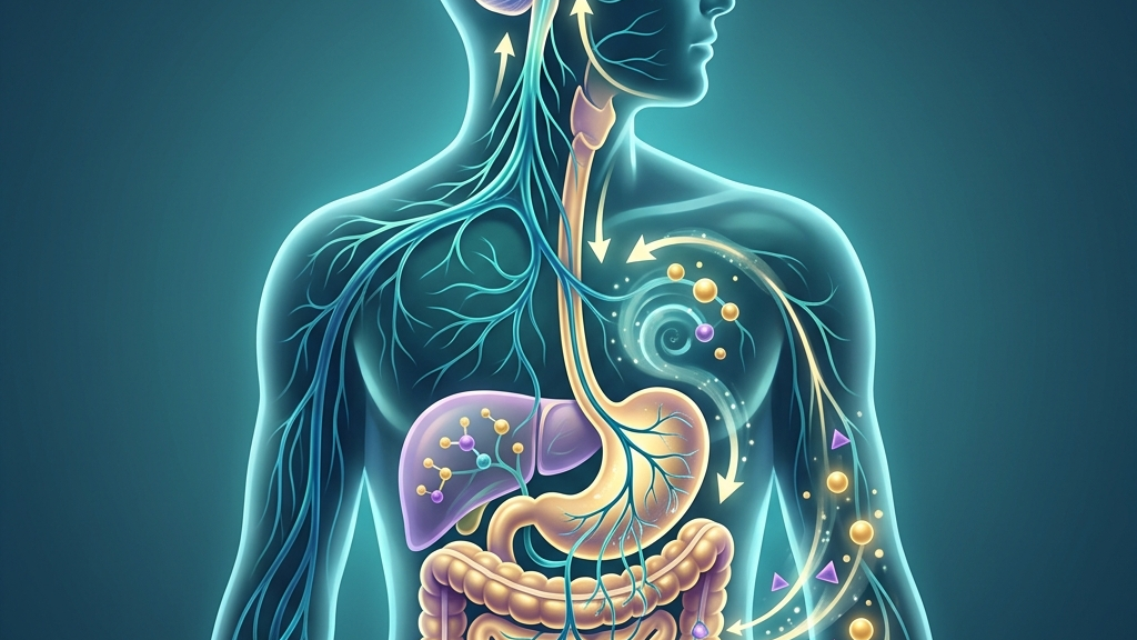
The vagus nerve, designated as cranial nerve X, originates in the medulla oblongata and extends long branches that reach multiple thoracic and abdominal organs. Its parasympathetic fibers carry outgoing signals that promote conservation and restoration, slowing heart rate, stimulating digestive secretions, and supporting intestinal motility. These efferent pathways are complemented by a large proportion of afferent fibers that return sensory information from the viscera to the brainstem, creating a closed loop of monitoring and adjustment. The efferent cell bodies reside primarily in the dorsal motor nucleus, whose axons travel through the vagal trunk and synapse onto postganglionic neurons embedded in the myenteric plexus; acetylcholine released at these synapses binds to muscarinic receptors on smooth muscle and secretory cells, raising intracellular calcium and thereby initiating both peristaltic contractions and glandular output. Meanwhile, the afferent cell bodies lie in the nodose and jugular ganglia, conveying mechanosensory, chemosensory, and nociceptive information via unmyelinated or thinly myelinated fibers that terminate in the nucleus tractus solitarius. Within the gut-brain axis, the vagus nerve transmits both mechanical and chemical signals generated by the stomach and intestines. Stretch receptors in the stomach wall, nutrient sensors in the mucosa, and microbial metabolites all feed information upward, allowing the brain to modulate digestive activity in real time. This bidirectional traffic helps coordinate hunger signals, satiety responses, and the timing of peristaltic waves. For instance, when a person eats a large mixed meal, progressive gastric distension activates tension-sensitive vagal endings that increase their firing rate proportionally to wall tension; this graded input reaches the hypothalamus and can suppress further appetite while simultaneously enhancing pyloric relaxation to allow controlled emptying. Chemical detection occurs through vagal fibers that express receptors for glucose, amino acids, and fatty-acid derivatives, permitting the brainstem to distinguish a carbohydrate-heavy breakfast from a protein-dominant dinner and to adjust downstream pancreatic and biliary responses accordingly. Heart-rate variability serves as one accessible window into vagal function. Greater variability between successive heartbeats often reflects stronger parasympathetic influence via the vagus, whereas reduced variability can indicate a shift toward sympathetic dominance. Researchers measure this marker because it correlates with the same brainstem nuclei that govern gastrointestinal tone, providing an indirect but practical index of overall vagal engagement. In practice, an individual seated at a desk who performs a brief sequence of slow diaphragmatic breaths may observe an immediate rise in the root-mean-square of successive differences between heartbeats; this change indexes heightened activity in the nucleus ambiguus, whose cardioinhibitory neurons share circuitry with those projecting to the gut. Over longer timescales, regular aerobic conditioning tends to elevate resting high-frequency heart-rate variability, reflecting structural and functional adaptations in vagal preganglionic neurons. The nerve’s extensive distribution also means that activity in one region can influence another. For instance, changes in respiratory rhythm directly affect vagal outflow to the heart and, simultaneously, to the enteric nervous system. This anatomical overlap explains why practices that alter breathing patterns frequently coincide with shifts in digestive comfort, although the precise pathways continue to be mapped in greater detail. During quiet exhalation, pulmonary stretch receptors and baroreceptors reinforce vagal cardioinhibitory tone; the same brainstem interneurons that receive this pulmonary input also modulate the dorsal motor nucleus, so that a slower respiratory cycle can entrain a more regular pattern of gastric migrating motor complexes. Conversely, rapid shallow breathing during periods of mental workload reduces pulmonary afferent drive and thereby withdraws a portion of vagal support from both cardiac and intestinal targets, illustrating the shared regulatory pool.Regulating Digestion Through Vagal Pathways
Vagal efferents release acetylcholine at their terminals within the enteric nervous system, directly stimulating smooth-muscle contractions that propel contents through the stomach and intestines. The same neurotransmitter promotes glandular secretion of acid, enzymes, and mucus, creating the chemical environment required for initial protein breakdown and protection of the mucosal lining. When vagal traffic is robust, these coordinated actions tend to proceed in orderly sequence; when traffic diminishes, motility and secretion can slow or become irregular. Acetylcholine also acts presynaptically on enteric interneurons to coordinate the timing of ascending excitatory and descending inhibitory reflexes that underlie peristalsis, so that each wave of contraction is matched by appropriate relaxation ahead of the bolus. Afferent vagal fibers continuously report the degree of gastric distension, the presence of specific macronutrients, and local inflammatory signals back to the nucleus tractus solitarius. This sensory stream allows the brain to fine-tune subsequent motor output, for example by increasing pancreatic enzyme release once fats are detected or by adjusting gastric emptying rate according to caloric density. Disruption anywhere along this afferent–efferent loop can alter the timing and completeness of digestion without necessarily involving structural damage. In an everyday setting, someone who consumes a high-fat meal while under time pressure may experience delayed gastric emptying because concurrent sympathetic activation overrides vagal facilitation of pyloric relaxation; the same meal eaten after a short walk may empty more promptly because residual vagal tone remains available to integrate the lipid-induced afferent signals. Many people notice that meals eaten during rushed or emotionally charged moments leave them with sensations of heaviness or incomplete processing hours later. Conversely, the same foods consumed in quieter settings sometimes move through the tract with less awareness of discomfort. These subjective differences align with known shifts in vagal tone that accompany changes in arousal level, although individual variability in baseline tone and concurrent factors such as posture and hydration also contribute. Postprandial recordings of gastric myoelectrical activity show that sympathetic arousal reduces the proportion of normal three-cycles-per-minute slow waves, whereas relaxed conditions preserve this rhythm and allow vagal efferents to maintain orderly propagation. The vagus further participates in the migrating motor complex, the cyclical pattern of contractions that clears residual material from the stomach and small intestine between meals. Adequate vagal signaling supports the initiation and propagation of these waves, which in turn influences microbial balance and the sensation of morning appetite. When vagal modulation is less consistent, the intervals between these cleansing contractions may lengthen, an observation reported across multiple studies of gastrointestinal motility. The phase-III contractions of the migrating motor complex are preceded by a rise in vagal efferent discharge that coincides with peaks in motilin and ghrelin; this coordinated neurohormonal event sweeps undigested residue and sloughed epithelial cells distally, limiting bacterial overgrowth in the proximal small bowel.What the Research Shows
Studies mapping the anatomic course of the vagus have clarified how its branches interface with both the myenteric and submucosal plexuses, providing a structural basis for the motility and secretory effects described above. Neuroanatomy, Cranial Nerve 10 (Vagus Nerve) outlines the mixed afferent–efferent composition that enables this continuous feedback. Additional tracing studies reveal that vagal axons form dense arborizations around ganglia in the antrum and duodenum, positioning them to influence both the pacemaker interstitial cells of Cajal and the secretomotor neurons of the submucosal plexus. Research focused on the brain–gut axis has demonstrated that vagal sensory neurons respond to microbial metabolites and inflammatory cytokines, transmitting these signals centrally and thereby influencing both digestive reflexes and mood-related circuits. Vagus Nerve as Modulator of the Brain–Gut Axis reviews the evidence that selective vagotomy or stimulation alters gut permeability and visceral sensitivity in experimental models. In these preparations, transection of vagal trunks eliminates the ability of certain bacterial strains to modulate anxiety-like behavior, underscoring the nerve’s role as a conduit for microbiota-derived information. Additional work has examined vagal sensory neurons specifically, showing that distinct subpopulations encode mechanical stretch versus nutrient composition, allowing graded control over gastric emptying and intestinal transit. Vagal Sensory Neurons and Gut–Brain Signaling details how these neuron classes contribute to the integrated control of digestion. Optogenetic activation of the stretch-sensitive population reliably delays gastric emptying, whereas selective stimulation of nutrient-responsive fibers accelerates pancreatic secretion without altering wall tension. Heart-rate variability measurements have been used to index cardiac vagal tone and, by extension, the broader parasympathetic state that supports gastrointestinal function. Heart Rate Variability and Cardiac Vagal Tone discusses methodological considerations and the physiological interpretation of these metrics in relation to visceral regulation. Frequency-domain analysis separating high-frequency power (0.15–0.40 Hz) from low-frequency components permits investigators to isolate parasympathetic contributions and to correlate them with simultaneous recordings of antroduodenal motility.Practical Ways to Support Your Vagus Nerve
- Slow, extended exhales performed for several minutes can increase parasympathetic outflow, which in turn may support the vagal signals that promote gastric motility and secretion.
- Humming or gentle gargling creates vibrations that travel along vagal branches innervating the throat and can be incorporated briefly after meals or during quiet moments of the day.
- Gentle cold exposure, such as splashing cool water on the face, engages vagal afferents and may produce a transient shift toward parasympathetic dominance without requiring extreme temperatures.
- Paced breathing at roughly six breaths per minute has been observed to raise heart-rate variability, a marker linked to the same brainstem centers that regulate digestive tone.
- Light movement such as walking after eating encourages mechanical and neural stimuli that complement vagal motor output to the intestines.
- Consistent morning light exposure together with a regular sleep schedule helps stabilize circadian rhythms that interact with vagal regulation of gastrointestinal function throughout the 24-hour cycle.
When to Talk to a Professional
Persistent difficulty swallowing, unexplained weight loss, or sudden changes in bowel habits warrant prompt medical evaluation regardless of any interest in nervous-system regulation. Severe or rapidly worsening abdominal pain, vomiting that prevents fluid retention, or rectal bleeding are additional signals that require clinical assessment rather than self-directed exploration. Individuals who experience fainting, chest pain, or marked fluctuations in heart rhythm should seek professional guidance before experimenting with breathing or cold-exposure practices. A qualified clinician can determine whether these symptoms stem from vagal or other physiological sources and can provide appropriate testing or referral.Common Questions
How quickly might vagal tone change with simple breathing practices?
Acute shifts in heart-rate variability can appear within minutes of slow breathing, yet sustained alterations in baseline tone generally require repeated practice over days to weeks, with considerable individual variation. The rapidity of the acute effect depends on the degree to which respiratory sinus arrhythmia is already entrained; individuals with higher resting vagal tone often show larger immediate gains, whereas those whose baseline tone is lower may require several sessions before the same magnitude of change is observed.
Does posture affect vagal signaling to the gut?
Upright or slightly reclined positions after eating tend to facilitate the mechanical aspects of digestion that vagal motor fibers help coordinate, whereas slouched postures may mechanically compress abdominal structures and indirectly influence motility patterns. Gravity-assisted bolus movement in the upright position reduces the work required of vagally mediated peristalsis, while forward flexion can increase intra-abdominal pressure and transiently alter the threshold for vagal afferent activation.
Can vagal afferents detect specific nutrients?
Yes, subpopulations of vagal sensory neurons respond preferentially to certain macronutrients and to mechanical stretch, allowing the brain to adjust subsequent secretory and motor responses accordingly. Electrophysiological recordings demonstrate that fibers innervating the proximal duodenum increase their discharge rate within seconds of lipid arrival, whereas glucose-sensitive fibers located more distally respond with a slower time course that matches the rate of absorption.
Is there a relationship between vagal function and microbial composition?
Experimental and observational data indicate bidirectional interactions in which vagal signaling influences gut motility and permeability, while microbial metabolites in turn activate vagal afferents that report to the brainstem. Motility patterns shaped by vagal efferents determine the residence time of luminal contents, thereby shaping the ecological niche available to different bacterial taxa; in the reverse direction, metabolites such as indole and butyrate bind to receptors on vagal terminals and modify afferent firing frequency.
The vagus nerve’s extensive reach into digestive physiology underscores the value of viewing gastrointestinal function as part of a larger, integrated regulatory system rather than an isolated process. Attention to the mechanisms that link brainstem activity, visceral sensation, and intestinal motility offers a grounded framework for understanding everyday digestive experience. Continued research will refine these connections, yet the existing anatomical and physiological evidence already supports a coherent picture of parasympathetic participation in gut regulation.Have a question?
Have a question about something specific? Send us a message.
Visit VagusSkool.com/contact — we'll try to get back to you within 24 hours.