The Vagus Nerve and Digestion: Mechanisms Linking Cranial Signaling to Gastrointestinal Function
By Sterling Cooley
•
June 7, 2026
The vagus nerve serves as a primary bidirectional pathway between the brainstem and the digestive tract, allowing the central nervous system to monitor and adjust gut activity in real time. This connection matters because digestive comfort, nutrient absorption, and motility patterns often reflect the degree of parasympathetic engagement rather than isolated organ problems. Understanding these links provides a clearer picture of why sensations such as fullness, bloating, or irregular transit can shift when overall nervous-system balance changes.
To appreciate the scale of this influence, consider how a single nerve can integrate signals across multiple organ systems. The vagus nerve carries information about the mechanical state of the stomach wall after a meal, the pH level inside the duodenum, and even the presence of certain microbial metabolites produced in the colon. These data streams converge in the brainstem, where they are compared against current metabolic needs and emotional context before new instructions are sent back down the same fibers. Because the same nuclei also govern cardiac rhythm and respiratory depth, a shift in digestive signaling can subtly alter heart-rate patterns within the same minute, illustrating the integrated nature of autonomic control.
Readers will encounter the anatomical layout of the vagus nerve, its specific contributions to gastrointestinal motility and secretion, and the physiological observations that frequently accompany altered vagal signaling. Evidence from neuroanatomy and physiological studies will be examined without implying clinical outcomes. The discussion closes with everyday practices that some individuals explore to support vagal pathways and guidance on when professional evaluation is appropriate.
How the Vagus Nerve Works
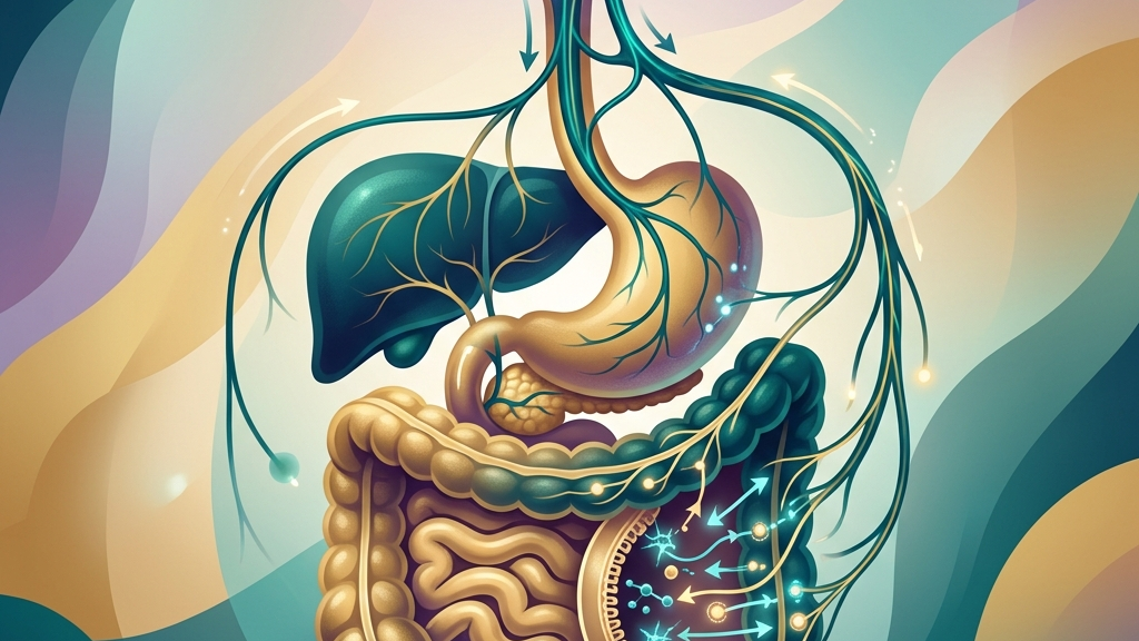
The vagus nerve, designated as cranial nerve X, originates in the medulla oblongata and extends long branches that reach the esophagus, stomach, small intestine, and proximal colon. Roughly eighty percent of its fibers are afferent, carrying sensory information from visceral organs back to the brainstem, while the remaining efferent fibers deliver parasympathetic instructions that promote rest-and-digest states. This arrangement positions the nerve as the main conduit of the parasympathetic division, counterbalancing sympathetic activation during periods of safety and recovery. The anatomical path begins with cell bodies clustered in the nodose and jugular ganglia just outside the skull base. From there, the nerve descends through the neck alongside the carotid artery, gives off branches to the larynx and pharynx, and then passes through the diaphragm to enter the abdominal cavity. Once inside the abdomen, anterior and posterior trunks form a loose plexus around the esophagus before fanning out to individual organs. This extensive distribution means that a single brainstem decision can influence esophageal sphincter tone, gastric acid output, and small-bowel segmentation almost simultaneously. Within the gut-brain axis, vagal afferents detect mechanical stretch, chemical composition of chyme, and local immune signals, then relay these data to the nucleus tractus solitarius. From there, processed information influences hypothalamic and limbic centers that modulate appetite, mood, and autonomic outflow. Heart-rate variability serves as a peripheral window into this traffic; higher variability, particularly in the high-frequency band, often corresponds to stronger vagal cardio-inhibitory tone and is interpreted by researchers as an index of parasympathetic flexibility. Because the same brainstem nuclei coordinate both cardiac and gastrointestinal efferents, changes in vagal tone can appear simultaneously in heart rhythm and digestive timing. When vagal outflow increases, gastric pacemaker cells receive more consistent excitatory input, and intestinal smooth muscle tends to maintain rhythmic segmentation. Conversely, reduced vagal engagement may coincide with slower gastric emptying or diminished pancreatic enzyme release, illustrating the nerve’s integrative rather than isolated role. In everyday terms, someone who notices their pulse slow after sitting down to a relaxed meal is experiencing the same brainstem shift that also lengthens the interval between antral contractions, allowing more thorough mixing of food with gastric juices.How the Vagus Nerve Orchestrates Digestive Processes
Vagal efferent fibers synapse onto enteric neurons throughout the myenteric and submucosal plexuses, releasing acetylcholine that excites smooth-muscle contraction and stimulates glandular secretion. In the stomach, this input drives receptive relaxation of the fundus and coordinates antral peristalsis, allowing measured accommodation of food volume followed by controlled emptying into the duodenum. The same fibers also promote mucus and bicarbonate output from surface epithelial cells, contributing to mucosal defense during active digestion. Sensory vagal endings distributed along the gastrointestinal tract register distension and nutrient presence, triggering reflex adjustments via the brainstem. For example, duodenal chemoreceptors signal the arrival of lipids or amino acids, prompting a vagally mediated reduction in gastric tone that slows further emptying—an adaptive brake that prevents overwhelming the small intestine with undigested material. These reflexes operate largely below conscious awareness yet shape the timing and comfort of postprandial sensations. A concrete illustration occurs after eating a fatty meal: the sensation of prolonged fullness arises partly because vagal afferents have informed the brainstem that lipid digestion will require extra time, resulting in a temporary decrease in antral pump strength. Many people notice that digestive regularity or ease of swallowing varies with overall stress load. Periods of sustained sympathetic dominance can coincide with sensations of early satiety, sluggish transit, or variable bowel patterns, while intervals of greater calm often align with more predictable hunger cues and smoother motility. These subjective shifts do not prove causation but reflect the vagus nerve’s position as a shared regulator of both visceral tone and autonomic state. Consider the common experience of eating quickly during a work deadline: the accompanying sympathetic activation reduces the amplitude of vagal efferent bursts, so the lower esophageal sphincter may relax less completely and the stomach may accommodate less volume before signaling fullness. The enteric nervous system retains considerable autonomy, yet vagal input provides an overlay that fine-tunes amplitude and coordination across long distances of gut tube. When vagal traffic is robust, migrating motor complexes during fasting appear more organized, supporting the housekeeping contractions that clear residual contents. Diminished vagal contribution may leave these complexes fragmented, an observation sometimes linked by researchers to altered inter-meal intervals or gas accumulation. In practical terms, someone who skips breakfast after a late night may experience more audible intestinal sounds later in the morning because the usual vagally timed cleansing waves have been delayed.What the Research Shows
Neuroanatomical tracing studies confirm that vagal sensory neurons terminate in discrete mucosal and muscular layers of the gut wall, forming the structural basis for real-time monitoring of luminal contents. Vagal Sensory Neurons and Gut–Brain Signaling details how these neurons encode both mechanical and chemical stimuli and transmit them centrally within milliseconds, enabling rapid reflex adjustments in motility and secretion. Functional investigations of the brain–gut axis further illustrate that electrical or pharmacological enhancement of vagal efferents can modulate gastric compliance and intestinal transit in animal models. Vagus Nerve as Modulator of the Brain–Gut Axis reviews evidence that vagal pathways integrate immune and microbial signals from the gut with brainstem autonomic centers, suggesting a route by which peripheral inflammation may influence central drive to the gastrointestinal tract. Human physiological recordings using heart-rate variability metrics show associations between higher vagal tone and more stable inter-beat intervals during rest. Heart Rate Variability and Cardiac Vagal Tone discusses how these metrics correlate with parasympathetic outflow that also reaches the gut, although direct causation for specific digestive patterns remains under study. Cleveland Clinic summaries likewise note the vagus nerve’s extensive gastrointestinal distribution without attributing isolated symptom relief to any single maneuver. Collectively, these sources emphasize the nerve’s role as an information highway rather than a simple on–off switch. Variability in vagal responsiveness appears influenced by age, fitness, and concurrent inflammation, underscoring why population-level findings do not translate uniformly to individual experience. For instance, older adults often exhibit lower high-frequency heart-rate variability at rest, yet the same individuals may still demonstrate robust postprandial gastric relaxation if other lifestyle factors support vagal traffic.Practical Ways to Support Your Vagus Nerve
- Slow, extended exhales—lengthening the out-breath to roughly twice the duration of the in-breath—engage pulmonary stretch receptors that increase vagal cardio-inhibitory signaling and may indirectly influence gastric relaxation.
- Gentle humming or soft gargling creates mechanical vibration along the pharyngeal branches of the vagus, providing afferent stimulation that some individuals report accompanies a calmer abdominal sensation after meals.
- Brief, tolerable cold exposure such as cool water on the face activates the diving reflex, a vagally mediated response that can shift autonomic balance toward parasympathetic predominance within seconds.
- Paced diaphragmatic breathing at approximately six breaths per minute has been shown in laboratory settings to raise high-frequency heart-rate variability, a marker linked to broader vagal engagement that reaches gastrointestinal targets.
- Light rhythmic movement such as walking after eating promotes mechanical mixing in the gut while simultaneously elevating vagal tone through baroreceptor feedback from modest cardiovascular demand.
- Consistent morning light exposure paired with stable sleep timing helps entrain circadian oscillators that in turn regulate vagal outflow rhythms, supporting more predictable daily patterns of digestive motility.
When to Talk to a Professional
Sudden, severe abdominal pain, persistent vomiting, unexplained weight loss, or blood in the stool warrant prompt medical assessment regardless of any vagal considerations. These signs may indicate structural or inflammatory conditions that require diagnostic evaluation beyond autonomic regulation. Chronic or progressive changes in swallowing, bowel frequency, or postprandial comfort that interfere with daily function also merit professional review. A clinician can determine whether further testing for motility disorders, mucosal integrity, or systemic contributors is indicated.Common Questions
Does stimulating the vagus nerve directly improve digestion?
Research describes associations between vagal pathways and gastrointestinal motility, yet no specific stimulation technique has been shown to treat digestive disorders. Individual responses vary and should be discussed with a qualified practitioner.How long does it take to notice changes in vagal tone?
Heart-rate variability can shift measurably within minutes of certain breathing practices, but sustained alterations in digestive patterns typically unfold over longer periods and depend on multiple physiological factors.Are there risks to vagal maneuvers?
Most gentle practices such as slow breathing or humming carry minimal risk for healthy adults; however, individuals with cardiovascular conditions or swallowing difficulties should consult a clinician before experimenting with new techniques.Can diet influence vagal signaling to the gut?
Nutrient composition affects vagal afferent firing through chemosensory endings, yet dietary changes alone do not constitute a targeted intervention for vagal function and require individualized guidance. The vagus nerve supplies a continuous, bidirectional channel that integrates brainstem oversight with enteric activity, offering one physiological substrate for the familiar link between internal state and digestive experience. Exploring this relationship through measured practices and appropriate professional input can help individuals develop a more informed relationship with their own regulatory systems.Have a question?
Have a question about something specific? Send us a message.
Visit VagusSkool.com/contact — we'll try to get back to you within 24 hours.