Breathing Rhythms and the Vagus Nerve: Mechanisms of Respiratory Influence on Autonomic Balance
How the vagus nerve works
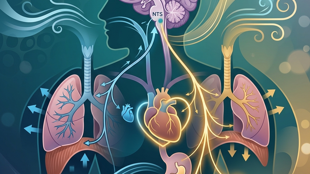
The vagus nerve, designated as cranial nerve ten, originates in the medulla oblongata and extends long branches through the neck, thorax, and abdomen. Its parasympathetic fibers slow heart rate, promote digestive secretions, and support recovery processes after stress. These fibers form part of the autonomic nervous system’s rest-and-digest arm, counterbalancing sympathetic activation during perceived threats or exertion. Mechanistically, preganglionic neurons in the dorsal motor nucleus and nucleus ambiguus send axons that synapse onto postganglionic neurons embedded in cardiac and gastrointestinal tissue; acetylcholine release at these synapses hyperpolarizes cardiac myocytes to lengthen the interval between beats and stimulates glandular cells to increase gastric acid and enzyme output. In daily life this appears when someone finishes a meal and notices a gradual slowing of heart rate alongside a sensation of abdominal fullness, reflecting coordinated vagal engagement that optimizes nutrient absorption while conserving energy. Beyond direct organ control, the vagus nerve participates in the gut-brain axis by transmitting sensory information from the gastrointestinal tract to brainstem nuclei. This bidirectional traffic influences mood-related circuits and can affect how the brain interprets signals of satiety or discomfort. Research on this pathway highlights its role in integrating visceral states with higher-order processing. Vagal afferents carry information about mechanical stretch in the stomach wall and chemical composition of chyme via specialized receptors; these signals reach the nucleus tractus solitarius and are relayed onward to hypothalamic and limbic areas that shape emotional tone. A concrete example occurs when an individual eats too quickly and later experiences both physical bloating and a subtle dip in mood, illustrating how afferent traffic can color cognitive appraisal of bodily comfort without conscious awareness of the neural route. Heart-rate variability, or the natural fluctuation in time between heartbeats, serves as one accessible window into vagal tone. Higher variability often correlates with stronger parasympathetic influence, while lower variability can appear during sustained sympathetic dominance. The vagus nerve contributes to this variability through its cardiac branches, which allow rapid adjustments in response to respiratory cycles and other inputs. The beat-to-beat changes arise because vagal efferent bursts are timed to the respiratory cycle, producing measurable differences in R-R intervals on an electrocardiogram. Someone monitoring a wearable device after a calm evening walk versus a tense work presentation may observe larger swings in interval length during the former, reflecting greater vagal modulation of the sinoatrial node under low-demand conditions.Breathing Rhythms and Vagal Tone
Respiration engages the vagus nerve through both mechanical and neural routes. During inhalation, lung stretch receptors and thoracic pressure changes stimulate vagal afferents that travel to the nucleus tractus solitarius. These signals can transiently reduce vagal outflow to the heart, producing the slight acceleration in heart rate observed in normal sinus arrhythmia. Exhalation reverses the pattern, allowing vagal tone to increase and heart rate to slow, creating the oscillatory pattern captured in heart-rate-variability metrics. The mechanical component involves phrenic and intercostal muscle activity that alters intrathoracic pressure, which in turn stretches baroreceptors in the aortic arch and carotid sinus; these receptors send additional afferent traffic along vagal pathways that fine-tune cardiac output on a breath-by-breath basis. The depth and pace of breathing further shape this dynamic. Slower, deeper breaths extend the expiratory phase, prolonging the period of heightened vagal cardiac inhibition and often elevating high-frequency components of heart-rate variability. Conversely, rapid or shallow breathing shortens expiratory windows and can reduce overall vagal modulation, a pattern sometimes observed during states of heightened alertness. These respiratory-vagal interactions occur continuously and adjust to metabolic demand without conscious effort. Consider a person transitioning from brisk walking to seated rest: the gradual lengthening of each exhale naturally augments vagal inhibition of the heart, visible as a progressive rise in interval variability on a simple pulse-oximeter display, whereas the same individual under time pressure may revert to quicker, upper-chest breaths that compress the expiratory window and blunt that variability. People commonly notice shifts in perceived calmness or bodily tension when breathing patterns change. Extended exhales, for instance, may coincide with a subjective sense of settling or reduced restlessness, consistent with increased vagal influence on heart rate and smooth muscle tone. Some individuals also report changes in digestive sensations or throat comfort during deliberate breathing practices, reflecting the nerve’s widespread distribution to visceral and laryngeal structures. The laryngeal motor fibers, for example, receive rhythmic input tied to expiratory lengthening, which can produce a subtle easing of throat muscle tension noticeable when someone hums softly while preparing breakfast. Individual responses vary with factors such as posture, fitness level, and concurrent emotional states. A person who habitually breathes with minimal diaphragmatic excursion may experience different baseline variability compared with someone whose breathing naturally incorporates fuller abdominal movement. These differences illustrate that respiratory patterns and vagal activity exist in a reciprocal relationship rather than a one-directional cause-and-effect sequence. An office worker who adopts a forward-head posture at a computer may reduce diaphragmatic range, thereby dampening the pressure swings that normally amplify vagal afferent traffic, while a trained singer accustomed to abdominal breathing may display more robust variability even during ordinary conversation.What the research shows
Studies using direct nerve recordings and non-invasive monitoring have mapped how respiratory phases align with fluctuations in vagal cardiac activity. Heart Rate Variability and Cardiac Vagal Tone reviews evidence that respiratory sinus arrhythmia arises largely from vagal modulation, providing a physiological basis for linking breathing rate to measurable autonomic indices. This work underscores that variability measures reflect integrated brainstem reflexes rather than isolated nerve firing. Complementary analyses of frequency-domain metrics show that the high-frequency band (0.15–0.40 Hz) predominantly indexes vagal contributions, allowing researchers to quantify how modest shifts in breathing rate move power within that band without invoking changes in sympathetic outflow. Investigations into sleep and respiration further illustrate vagal involvement. Vagus Nerve Stimulation, Sleep-Disordered Breathing & Sleep Quality examines how altered breathing during sleep can interact with vagal pathways, affecting both oxygen stability and autonomic recovery. The findings suggest that respiratory disruptions may dampen typical vagal surges that normally accompany restorative sleep stages. Overnight recordings reveal that slow-wave sleep normally coincides with pronounced expiratory lengthening and elevated vagal tone, whereas fragmented breathing reduces the duration of these periods and attenuates the associated rebound in heart-rate variability upon awakening. Additional research addresses the nerve’s sensory role in visceral signaling. Vagus Nerve as Modulator of the Brain–Gut Axis synthesizes data on how vagal afferents convey gut-derived information that can influence brainstem and forebrain circuits. Complementary work in Vagal Sensory Neurons and Gut–Brain Signaling details the molecular identities of these neurons and their contribution to interoceptive feedback loops that intersect with respiratory control centers. These loops allow nutrient sensing in the intestine to modulate respiratory rhythm generators in the medulla, providing a pathway through which a heavy meal can subtly slow breathing rate even before conscious satiety registers. Anatomical and clinical overviews from Vagus Nerve: Function, Location & Conditions and Neuroanatomy, Cranial Nerve 10 (Vagus Nerve) provide foundational descriptions of fiber distribution and brainstem nuclei that support the respiratory-autonomic connections observed in the studies above. Together these sources indicate consistent physiological linkages while noting that many functional outcomes remain subject to contextual and individual factors. Histological tracing demonstrates that approximately 80 percent of vagal fibers are afferent, underscoring why sensory feedback from lungs and viscera can rapidly recalibrate efferent tone to the heart within a single respiratory cycle.Practical ways to support your vagus nerve
- Slow extended exhales can be practiced by gently lengthening the out-breath relative to the in-breath, which aligns with natural increases in vagal cardiac modulation during expiration.
- Humming or gargling produces vibrations that travel along vagal motor fibers to the larynx and pharynx, offering a simple mechanical stimulus many people incorporate while performing routine hygiene tasks.
- Gentle cold exposure, such as splashing cool water on the face, engages trigeminal and vagal afferents that can shift autonomic balance toward parasympathetic predominance in some contexts.
- Paced breathing at approximately six breaths per minute has been examined for its effects on heart-rate variability and may be explored gradually by counting or using a simple timer.
- Light movement that coordinates breath with motion, such as walking while maintaining an easy rhythm, can integrate respiratory and vagal signaling without requiring intense effort.
- Morning light exposure combined with consistent sleep timing supports circadian regulation that indirectly influences autonomic tone, including vagal contributions to overnight recovery.
When to talk to a professional
Sudden or severe changes in heart rhythm, breathing difficulty at rest, or unexplained fainting warrant prompt medical evaluation regardless of any breathing practices being explored. Persistent digestive distress, chronic throat pain, or significant alterations in voice quality also merit professional assessment to rule out structural or neurological issues affecting vagal pathways. These symptoms may reflect compression, inflammation, or demyelination along the nerve’s long course rather than transient respiratory patterns, and only targeted examination such as laryngoscopy or autonomic function testing can distinguish among possibilities. Individuals experiencing symptoms that interfere with daily functioning, such as ongoing dizziness, profound fatigue, or mood changes accompanied by physical signs, should consult a qualified clinician. Self-observation of breathing patterns can be informative, yet it does not replace diagnostic procedures when symptoms suggest possible underlying conditions. Clinicians often integrate wearable data with laboratory measures of inflammatory markers or gastric emptying studies to build a comprehensive picture before attributing changes solely to breathing mechanics.Common questions
Does everyone experience the same breathing-related changes in vagal activity?
Responses vary with age, fitness, posture, and health status; some people show pronounced heart-rate variability shifts with breathing adjustments while others exhibit more modest changes, reflecting natural physiological diversity. For example, older adults may display reduced high-frequency power even at identical breathing rates because baroreflex sensitivity declines with vascular stiffening, whereas endurance athletes often maintain larger swings due to enhanced stroke volume and stronger vagal efferent traffic.
Can breathing practices influence inflammation through the vagus nerve?
Research suggests vagal pathways can modulate certain inflammatory signals, yet the magnitude and consistency of effects from breathing alone remain under investigation and differ across individuals. The cholinergic anti-inflammatory pathway involves vagal efferents that inhibit splenic macrophage cytokine release via nicotinic receptors, but human studies show wide inter-subject variability linked to baseline vagal tone and concurrent medication use.
How long do breathing-related shifts in heart-rate variability typically last?
Immediate changes often coincide with the duration of the altered breathing pattern, while longer-term adaptations may relate to repeated practice and overall lifestyle factors rather than single sessions. A single five-minute session of slowed breathing may elevate high-frequency power for roughly the same interval afterward, whereas weeks of consistent practice can shift resting respiratory rate itself, producing more sustained elevations in baseline variability.
Is there a single ideal breathing rate for vagal engagement?
No universal rate applies to all people; rates around six breaths per minute appear in studies for their association with variability increases, but comfort and sustainability guide individual exploration. Some individuals find that slightly faster rates still produce noticeable oscillatory effects when combined with adequate tidal volume, while others require slower tempos to avoid air hunger that itself activates sympathetic reflexes.
Do posture and movement affect how breathing influences the vagus nerve?
Yes, upright versus reclined positions and the presence of gentle movement alter diaphragmatic excursion and intrathoracic pressure, thereby modulating the mechanical and neural signals transmitted along vagal fibers. Supine positioning increases venous return and augments baroreceptor loading during expiration, amplifying vagal cardiac inhibition, whereas seated forward flexion can restrict lower rib movement and thereby attenuate the same pressure-mediated feedback. The interplay between breathing and vagal function illustrates one accessible route through which autonomic balance can be observed and, for many people, gently influenced over time. Continued attention to individual responses alongside professional guidance when needed supports an informed approach to these physiological relationships.
Have a question?
Have a question about something specific? Send us a message.
Visit VagusSkool.com/contact — we'll try to get back to you within 24 hours.